House of AnandaMovement · Stillness · Wisdom · Journey

New anatomy notes

Understand the skeleton, spine and joints.

Explore spinal curves, discs, nerve openings and the back-care notes alongside the existing bone-count and joint references.

Spine, discs & back care ↗ · Skull, ribs & spine ↗ · Joints & movement ↗

Muscle study · new notebook lesson

Understand contraction, not just stretching.

Compare muscle tissues and contraction types, then identify the neck, back, hip, hamstring and calf muscles using the existing labelled plates.

Muscles, contraction & movement ↗

House of Ananda · Anatomy for yoga

The body,
from head to toe.

A visual atlas of the skeleton, major muscle groups and body systems. Locate a structure, learn what it does, then connect it to movement and breath.

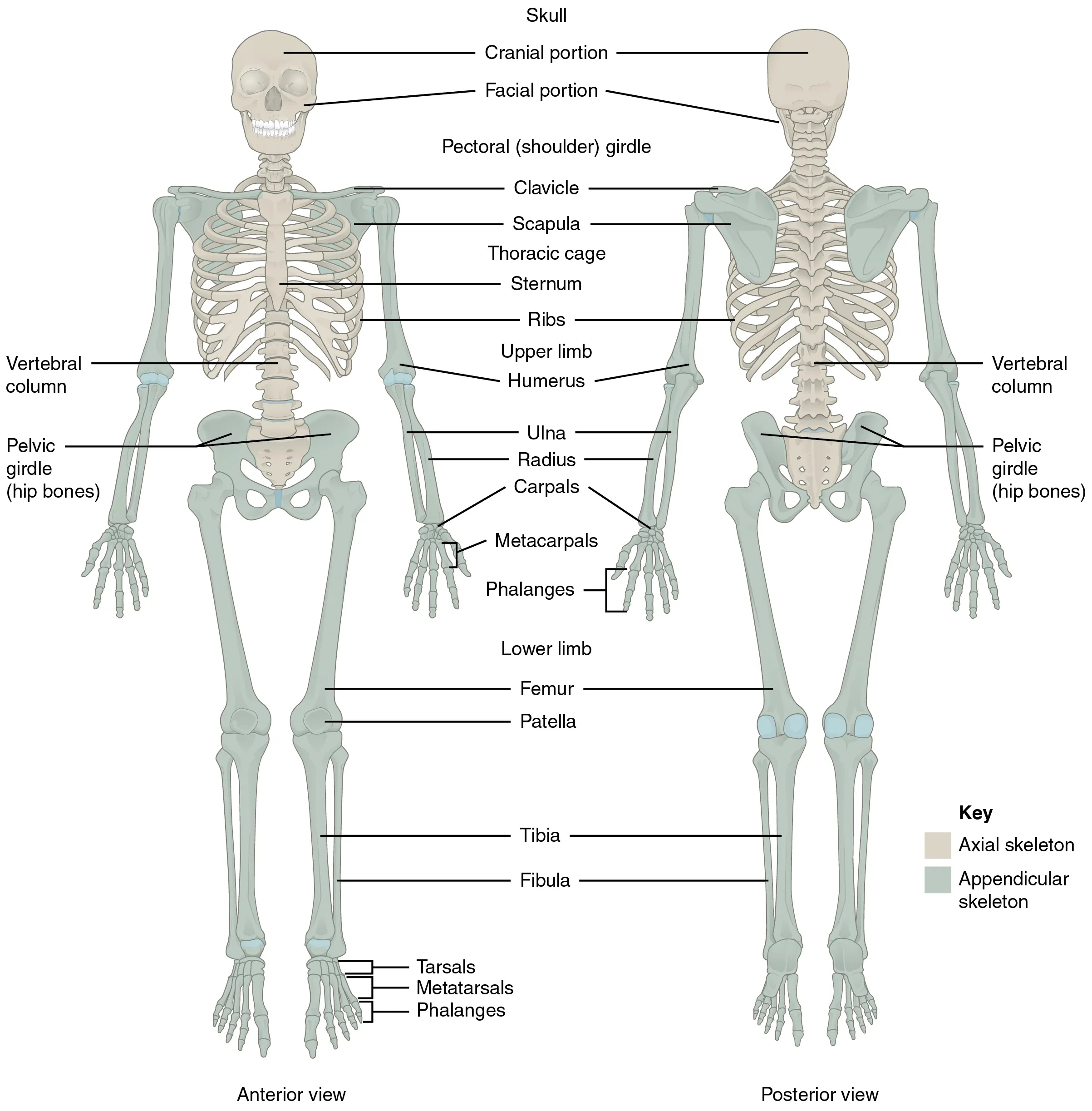
01 · The whole picture

Structure & movement

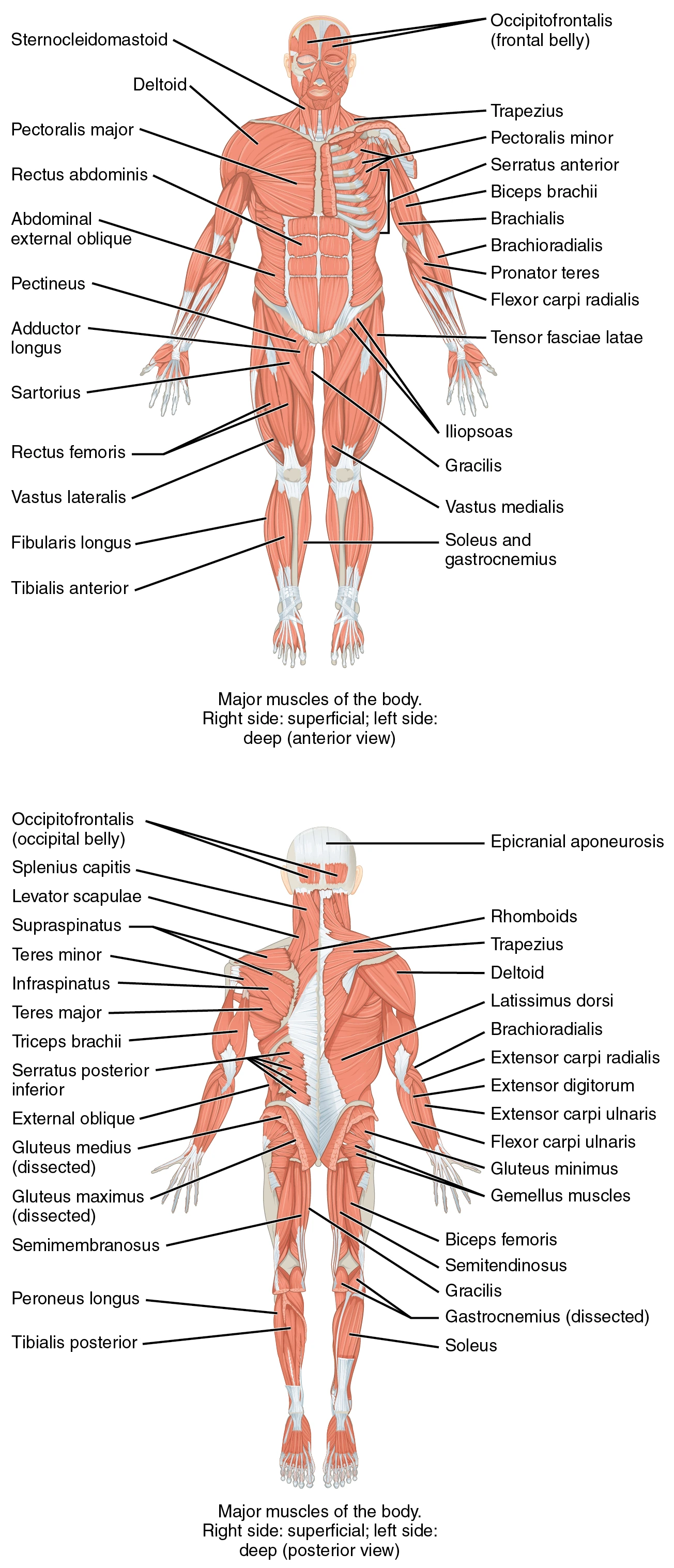
Front and back views show how the major bones and superficial muscles are arranged. Open any diagram at full size to read its labels.

Anatomical position means standing upright, facing forwards, with palms facing forwards. Left and right refer to the person shown. Cutaway illustrations remove selected layers to reveal deeper structures.

02 · The supporting framework

How the skeleton adds up

The standard adult count is 206 bones. Fusion and additional small bones vary between people; this is a learning convention.

80Axial
Head, spine & thorax
126Appendicular
Limbs & their girdles
206Standard adult total
Bone groups across the whole body
DivisionRegionBonesWhat is included
AxialSkull228 cranial + 14 facial bones
AxialEar ossicles & hyoid76 tiny middle-ear bones + 1 hyoid
AxialVertebral column2624 separate vertebrae + sacrum + coccyx
AxialThoracic cage2524 ribs + sternum
AppendicularShoulder girdles42 clavicles + 2 scapulae
AppendicularArms & hands6030 bones in each upper limb
AppendicularPelvic girdle22 hip bones
AppendicularLegs & feet6030 bones in each lower limb

Bone provides a framework and stores minerals.

Cartilage covers many joint surfaces and supports flexible structures.

Ligaments connect bones and help restrain joint movement.

Tendons transmit muscle force to bone.

divisions of the skeletal system ↗

03 · Study the regions

A closer look, head to toe

Each region pairs labelled plates with bone landmarks, muscle actions and a connection to yoga study.

Region 01

Head, jaw & neck

Orient the head. Soften unnecessary effort.

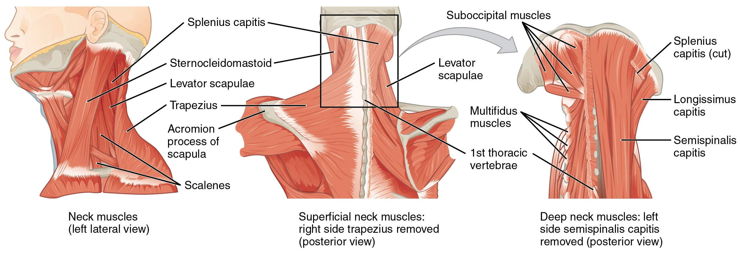
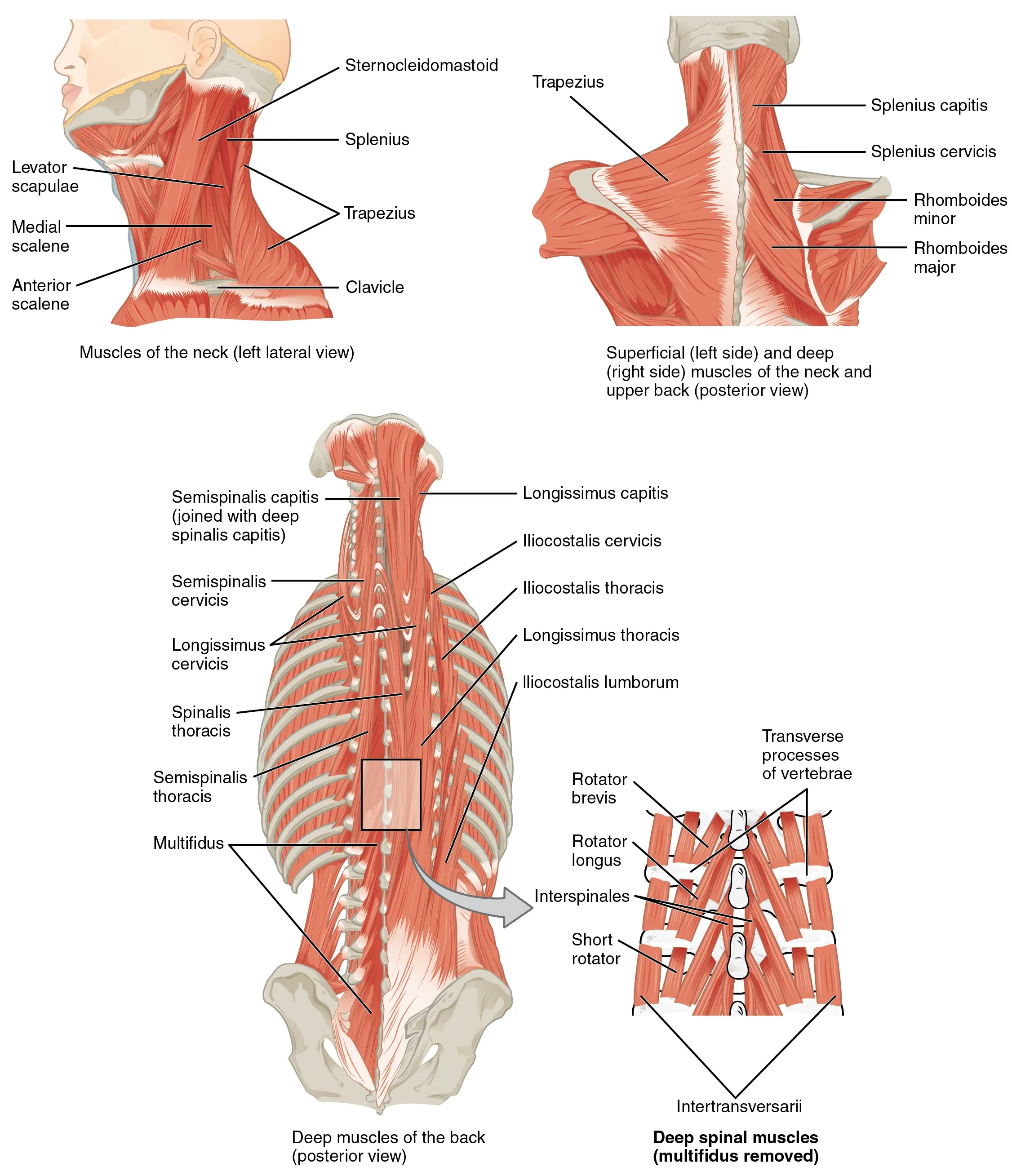
Labeled posterior and lateral views of superficial and deep neck muscles, including sternocleidomastoid, trapezius and splenius capitis.Open full-size diagram ↗
Muscles of the neckOpenStax · source · CC BY 4.0

Explore Pilates movement connections ↗

Bone landmarks

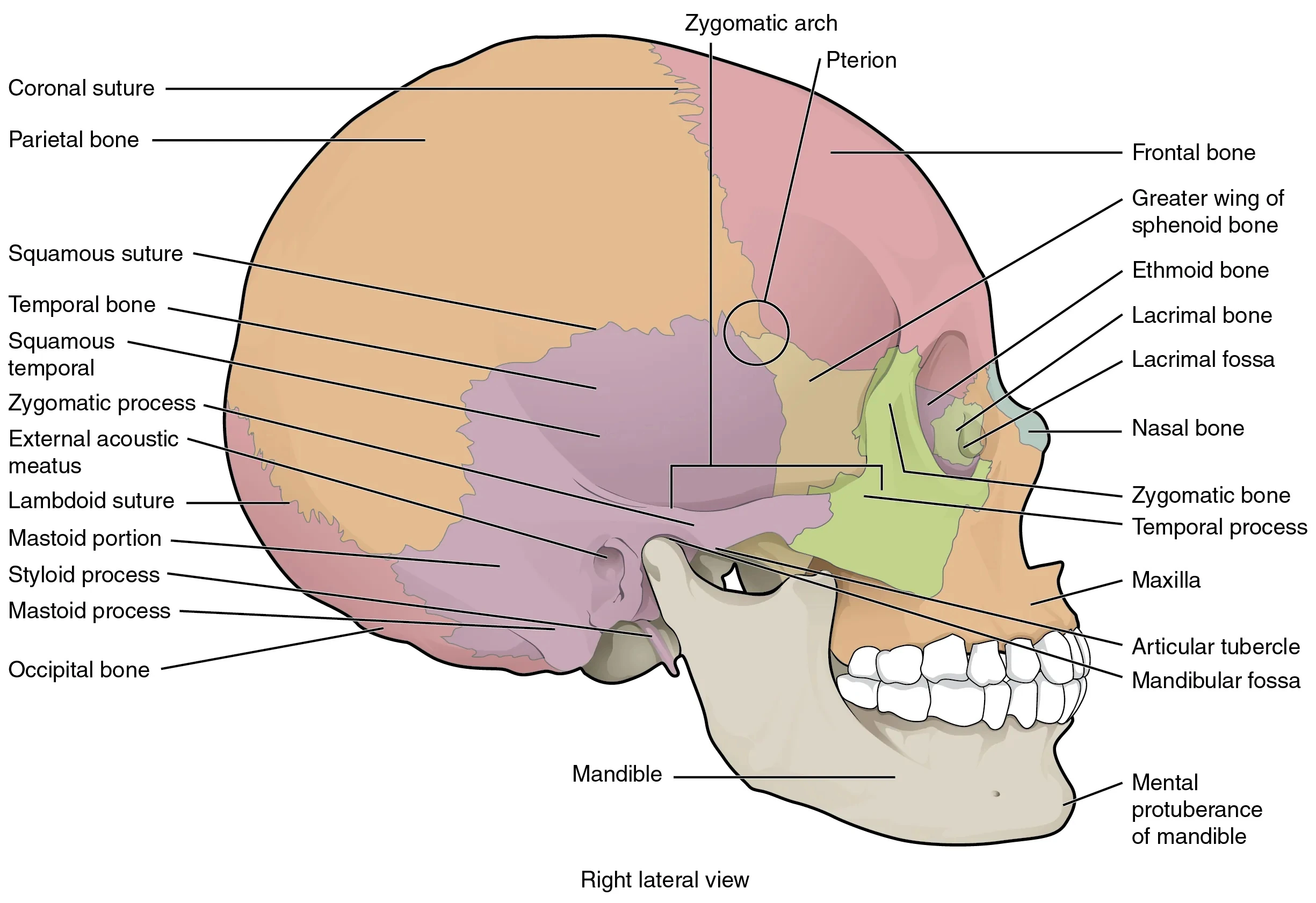
The skull includes the frontal, parietal, temporal, occipital, sphenoid and ethmoid bones around the brain, and facial bones including the maxillae, cheekbones and mandible. The hyoid lies in the neck. Seven cervical vertebrae support the head.

Sternocleidomastoid
Turns the head to the opposite side; can bend the neck.
Scalenes
Assist neck movement and can help elevate the upper ribs.
Masseter & temporalis
Close the jaw during chewing.
Deep neck muscles
Coordinate small adjustments of the head and cervical spine.
More diagrams · 1 plate

Connect to practice

In seated meditation, let your gaze and head position feel sustainable. A useful learning task is noticing jaw tension without pulling or forcing the neck.

Meditation foundations ↗
Back to body regions ↑

Region 02

Spine & back

A column of joints, supported by layers of muscle.

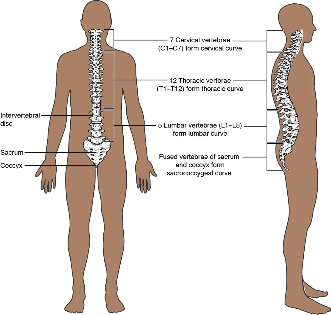
Labeled views of the vertebral column showing cervical, thoracic, lumbar, sacral and coccygeal regions and the spinal curves.Open full-size diagram ↗
The vertebral columnOpenStax · source · CC BY 4.0

Explore Pilates movement connections ↗

Bone landmarks

The movable spine has 7 cervical, 12 thoracic and 5 lumbar vertebrae. Below them sit the fused sacrum and coccyx. Discs between most vertebral bodies help distribute load; the spinal canal protects the spinal cord.

Erector spinae
Extend the spine and help control side bending.
Multifidus & other deep spinal muscles
Help control movement between spinal segments.
Quadratus lumborum
Assists side bending and supports the twelfth rib.
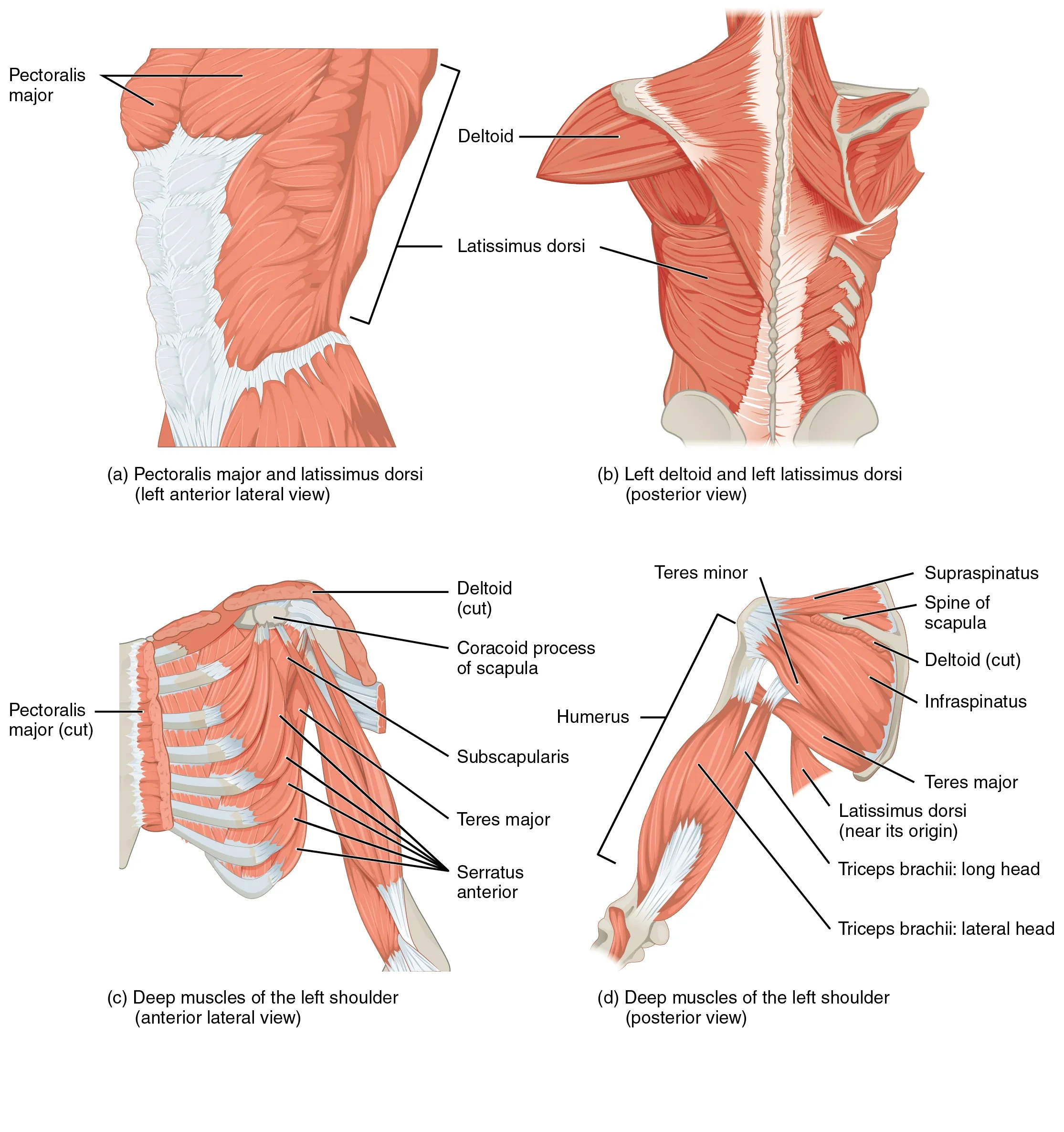
Latissimus dorsi
Links the trunk to the upper arm; draws the arm back and towards the body.
More diagrams · 1 plate

Connect to practice

Use the diagrams to distinguish a hip hinge from spinal flexion. Both occur in movement; the balance changes with the pose, the person and the task.

Pose library ↗
Back to body regions ↑

Region 03

Shoulders & upper back

The arm, shoulder blade and collarbone move together.

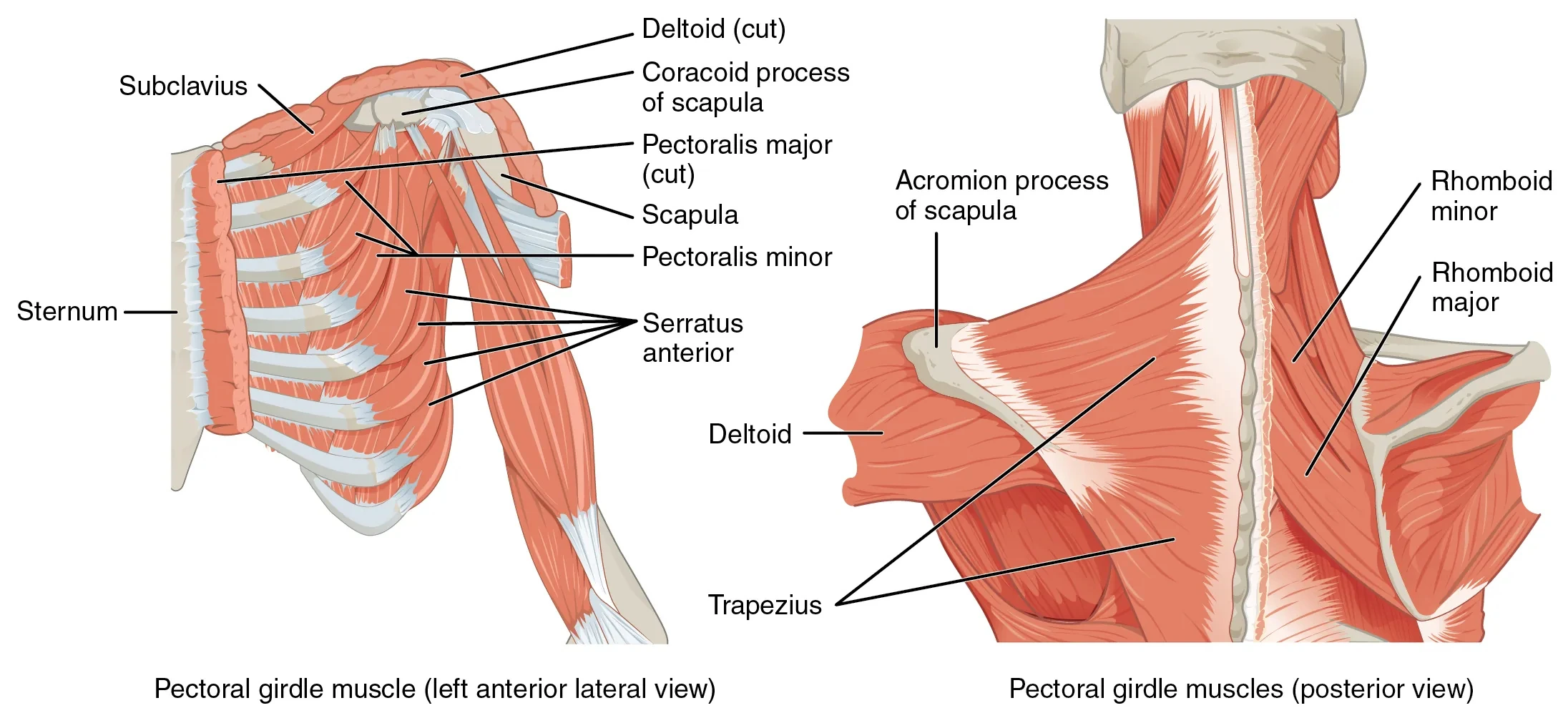
Anterior and posterior shoulder-girdle diagrams labeling trapezius, rhomboids, serratus anterior and other muscles that position the scapula.Open full-size diagram ↗
Muscles that position the shoulder girdleOpenStax · source · CC BY 4.0

Explore Pilates movement connections ↗

Bone landmarks

The clavicle is the collarbone; the scapula is the shoulder blade. The humeral head meets the scapula’s shallow socket. The acromion and scapular spine are useful landmarks in the diagrams.

Trapezius
Positions and stabilises the shoulder blade; its fibre groups contribute differently.
Serratus anterior
Draws the scapula around the ribs and helps it rotate upward.
Rhomboids
Draw the scapula towards the spine.
Rotator cuff
Supraspinatus, infraspinatus, teres minor and subscapularis help control the humeral head.
Deltoid
Lifts the arm; different portions contribute to different directions.
More diagrams · 1 plate

Connect to practice

An overhead reach involves movement of the shoulder blade as well as the arm. “Keep the shoulders down” is not a universal rule for every overhead pose.

Yoga series ↗
Back to body regions ↑

Region 04

Chest, ribs & breathing

A moving cage around the heart and lungs.

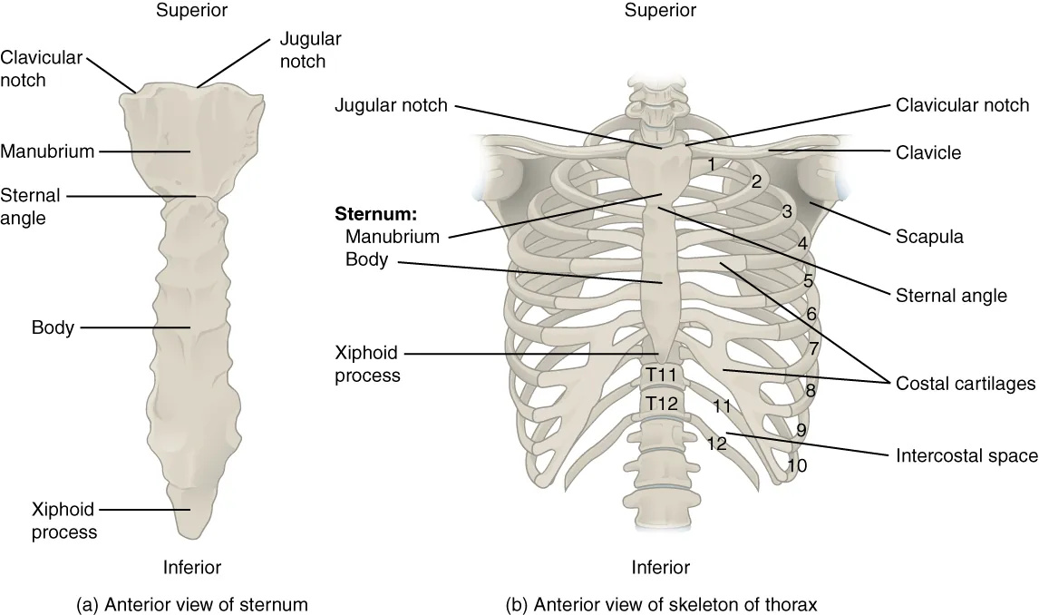
Labeled anterior views of the sternum and rib cage, showing the manubrium, sternal body, xiphoid process, ribs and costal cartilages.Open full-size diagram ↗
The rib cageOpenStax · source · CC BY 4.0

Explore Pilates movement connections ↗

Bone landmarks

The thoracic cage includes the sternum, 12 pairs of ribs and thoracic vertebrae. Costal cartilage links much of the rib cage to the breastbone; the lowest two pairs are floating ribs.

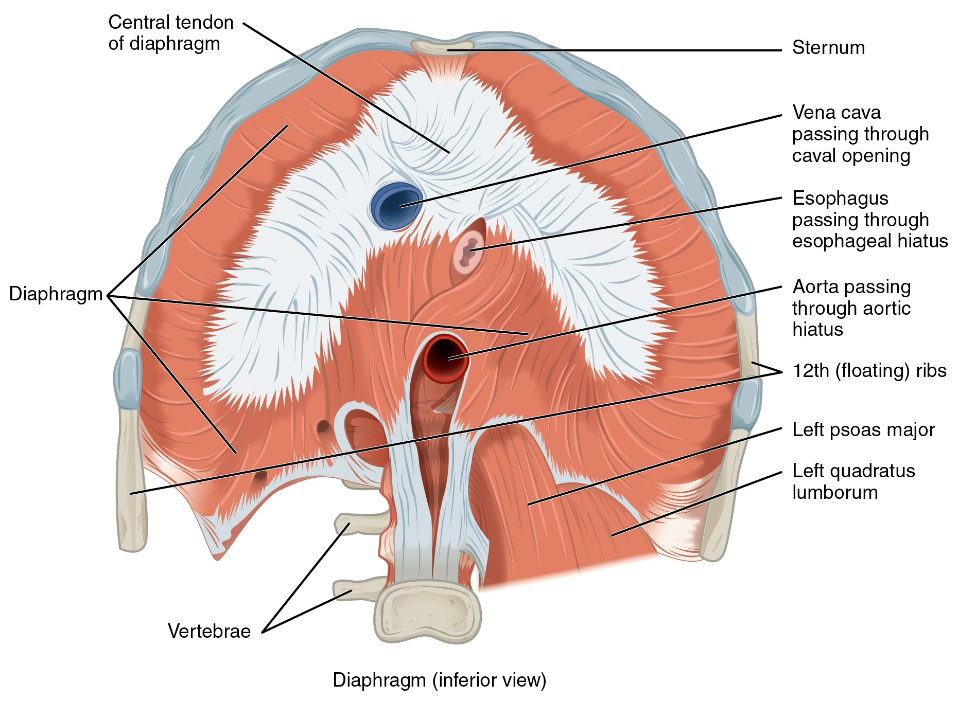
Diaphragm
Contracts and descends during inhalation, increasing chest-cavity volume.
Intercostals
Muscles between the ribs assist breathing mechanics.
Pectoralis major
Moves the upper arm across and towards the body.
More diagrams · 1 plate

Connect to practice

During quiet breathing, notice movement around the lower ribs. The lungs sit above the diaphragm; “belly breathing” describes visible abdominal movement, not air entering the abdomen.

Prāṇāyāma study ↗
Back to body regions ↑

Region 05

Abdomen, pelvis & pelvic floor

Support, pressure and movement through the centre.

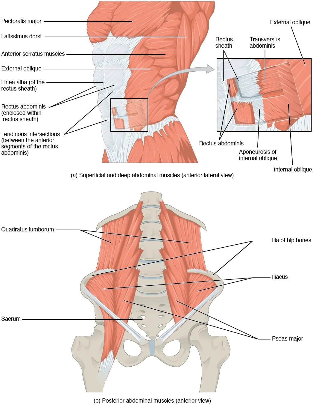
Labeled anterior and posterior abdominal-wall diagrams showing rectus abdominis, external and internal obliques, transversus abdominis and posterior abdominal muscles.Open full-size diagram ↗
Muscles of the abdomenOpenStax · source · CC BY 4.0

Explore Pilates movement connections ↗

Bone landmarks

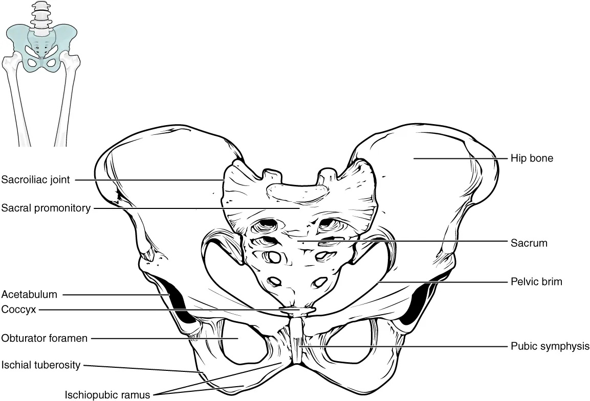
Each hip bone is formed from the fused ilium, ischium and pubis. Together with the sacrum and coccyx, the pelvic ring supports the trunk and connects it to the legs.

Rectus abdominis
Flexes the trunk.
Internal & external obliques
Contribute to trunk rotation and side bending.
Transversus abdominis
Contributes to abdominal compression and trunk support.
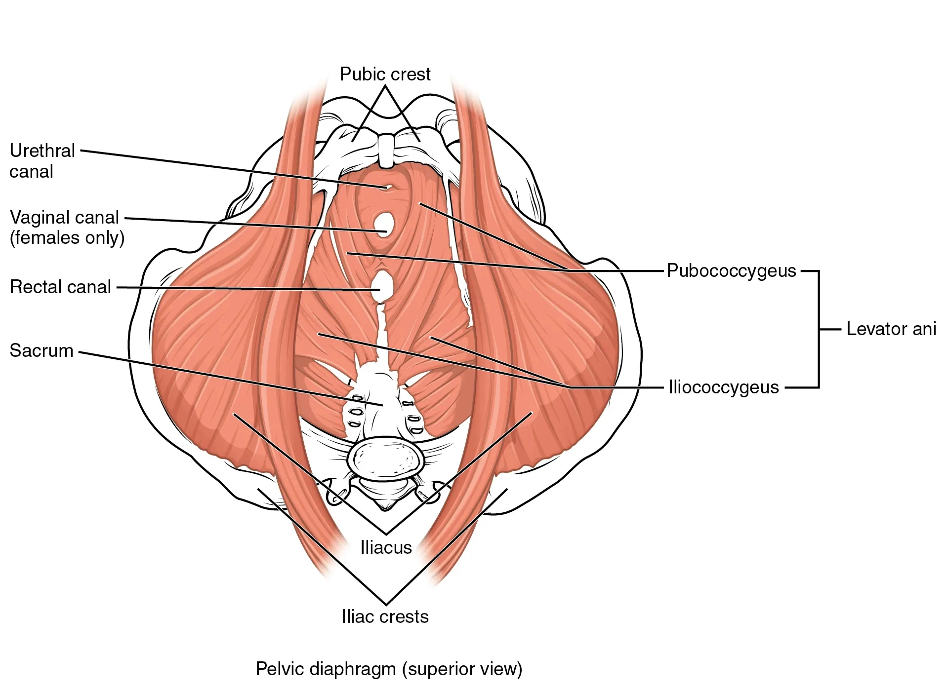
Pelvic floor
Supports pelvic organs and participates in continence and pressure control.
More diagrams · 2 plates

Connect to practice

The pelvic floor is not a single muscle. The anatomical diagrams are a guide to location; constant squeezing is not a general instruction for yoga or breathing.

Breath and stillness ↗
Back to body regions ↑

Region 06

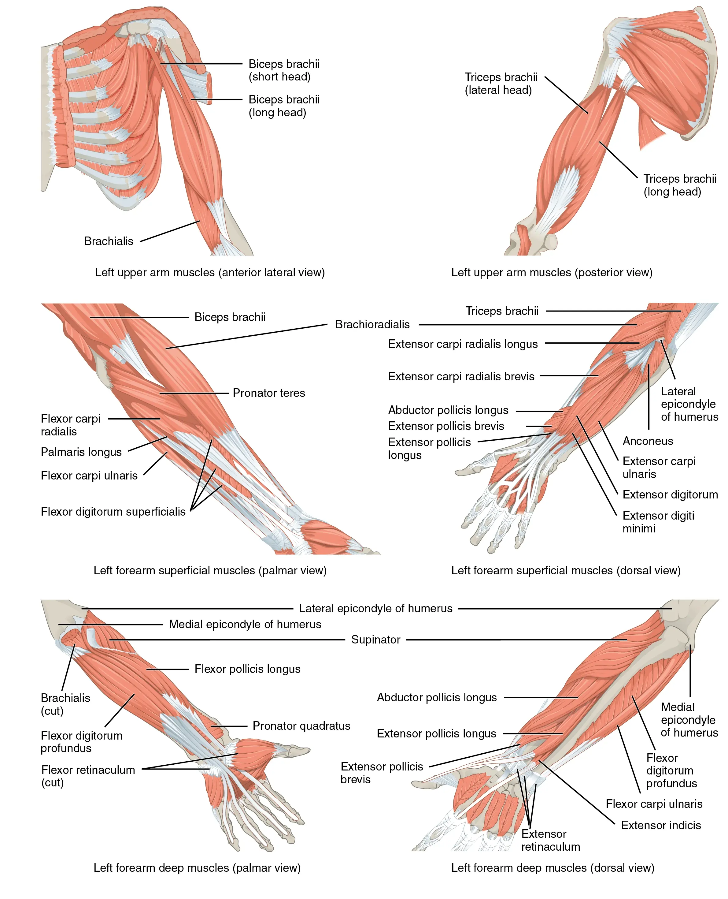
Upper arms & elbows

Bend, straighten and turn the forearm.

Labeled anterior and posterior views of upper-arm and forearm muscles, including elbow flexors and extensors and muscles acting on the wrist and fingers.Open full-size diagram ↗
Arm and forearm musclesOpenStax · source · CC BY 4.0

Explore Pilates movement connections ↗

Bone landmarks

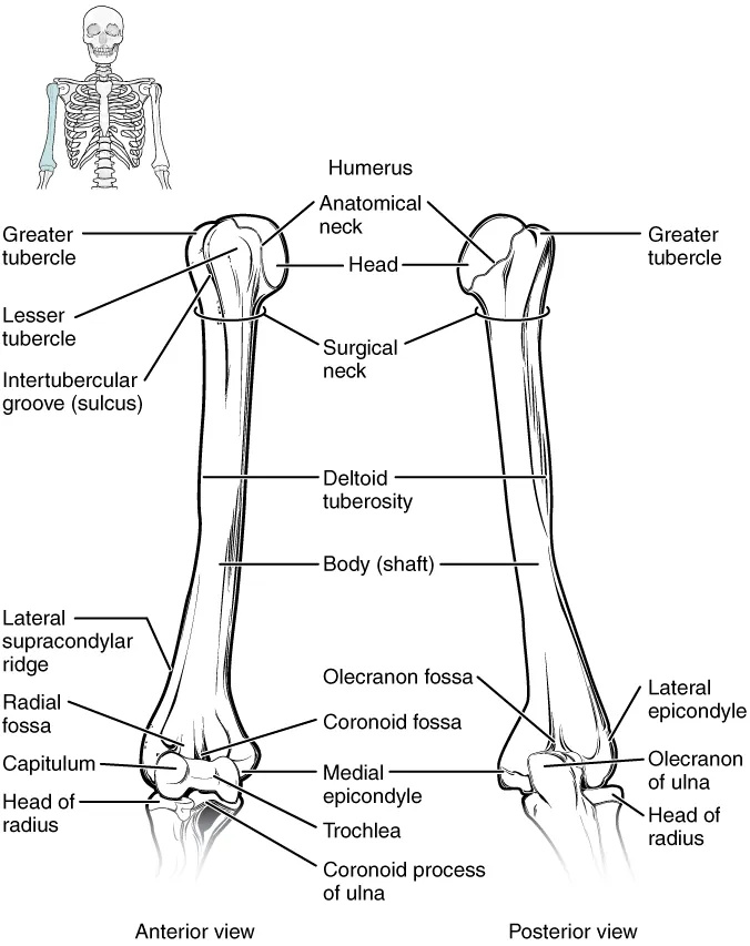
The humerus runs from shoulder to elbow. The radius lies on the thumb side of the forearm and the ulna on the little-finger side in anatomical position. They rotate relative to each other when the palm turns.

Biceps brachii
Bends the elbow and turns the palm upward.
Brachialis
Bends the elbow.
Triceps brachii
Straightens the elbow.
Pronators & supinator
Turn the forearm palm-down or palm-up.
More diagrams · 1 plate

Connect to practice

Compare arm positions in a wall-supported plank and a standing reach. Begin with a load that allows comfortable control rather than trying to copy a fixed shape.

Beginner flows ↗
Back to body regions ↑

Region 07

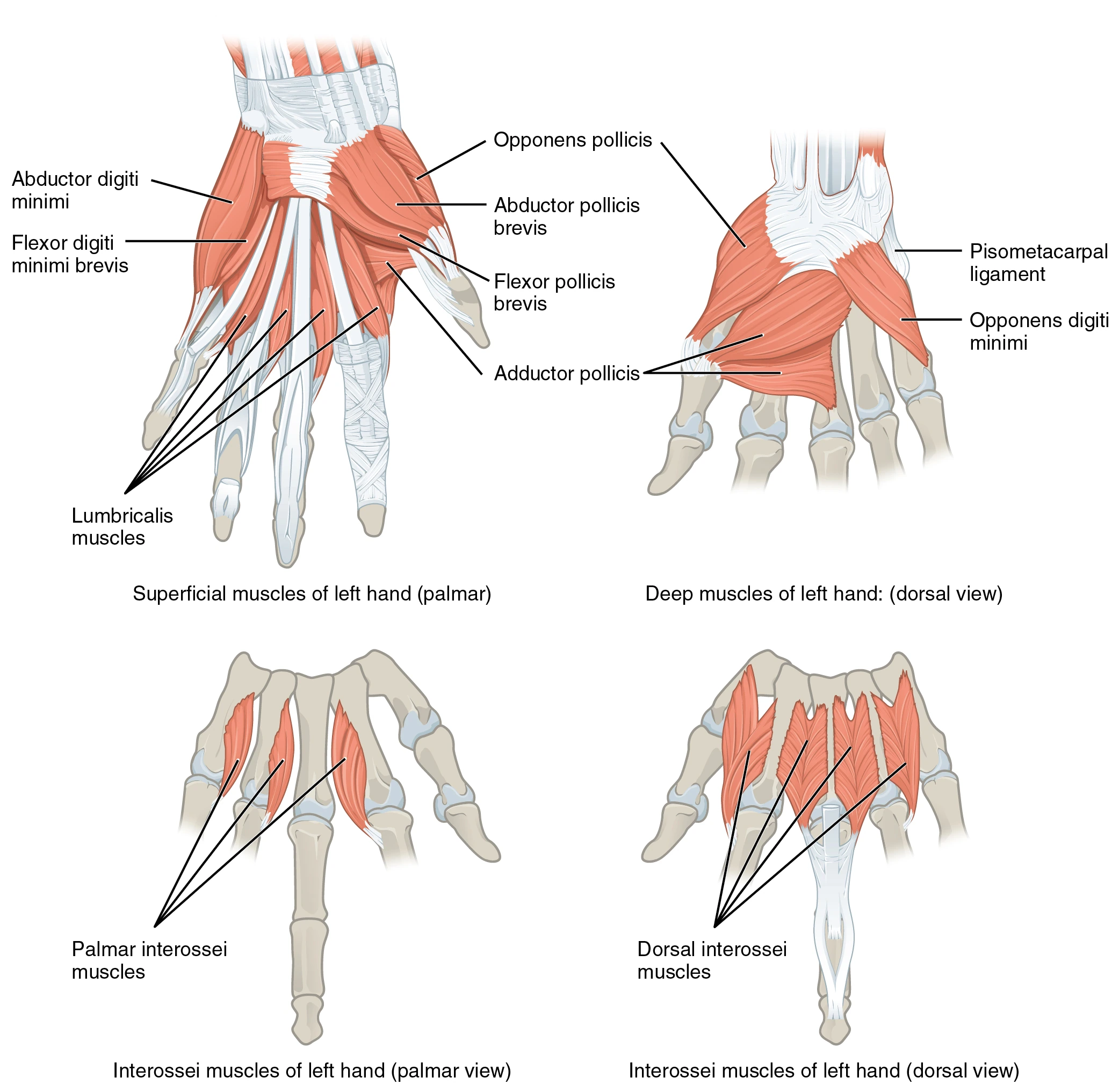
Forearms, wrists & hands

Small joints make both support and precision possible.

Labeled diagrams of superficial and deeper muscles within the hand, including thenar muscles, hypothenar muscles, lumbricals and interossei.Open full-size diagram ↗
Intrinsic muscles of the handOpenStax · source · CC BY 4.0

Explore Pilates movement connections ↗

Bone landmarks

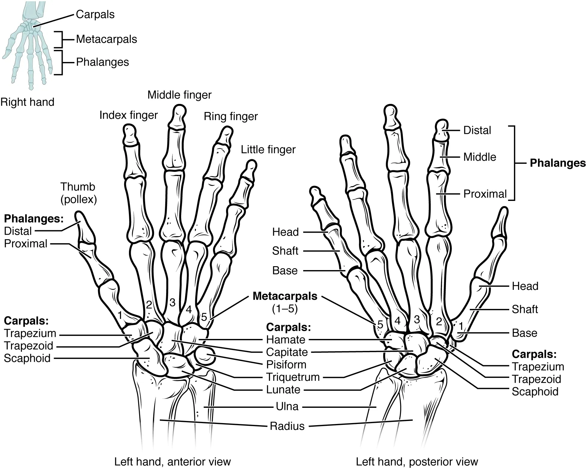
Each hand contains 8 carpals, 5 metacarpals and 14 phalanges. The carpals are the scaphoid, lunate, triquetrum, pisiform, trapezium, trapezoid, capitate and hamate. The thumb has two phalanges; each other finger has three.

Forearm flexors & extensors
Move the wrist and fingers through long tendons.
Thenar & hypothenar muscles
Control the thumb and little finger.
Interossei & lumbricals
Coordinate finger movement and fine control.
More diagrams · 1 plate

Connect to practice

A hand pose and a weight-bearing pose ask different things of the same anatomy. Explore comfortable finger positioning; increase load gradually when the palms support body weight.

Pose library and adaptations ↗
Back to body regions ↑

Region 08

Hips, buttocks & thighs

Large muscles move the femur and steady the pelvis.

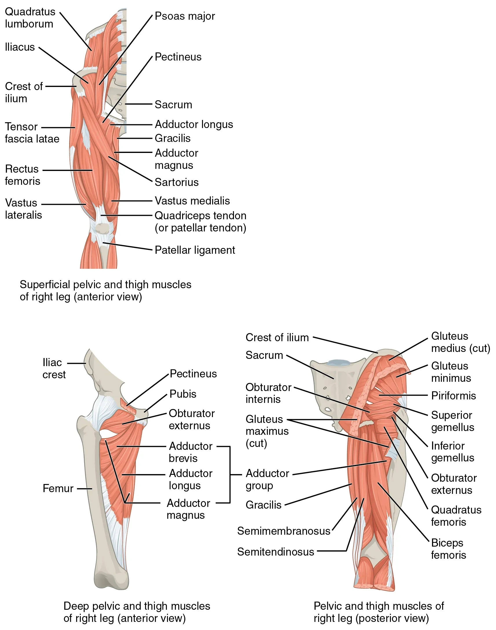
Labeled anterior and posterior views of the hip and thigh, showing gluteal muscles, quadriceps, hamstrings, adductors and other muscles acting on the hip and knee.Open full-size diagram ↗
Hip and thigh musclesOpenStax · source · CC BY 4.0

Explore Pilates movement connections ↗

Bone landmarks

The head of the femur sits in the acetabulum of the pelvis. The femoral neck and greater trochanter are landmarks near the hip. The hip is a ball-and-socket joint with individual differences in bone shape.

Iliopsoas
Psoas major and iliacus flex the hip.
Gluteal muscles
Gluteus maximus extends the hip; medius and minimus assist pelvic control and hip abduction.
Adductors
Draw the thigh towards the midline.
Deep hip rotators
Help control rotation of the femur.

Connect to practice

Hip rotation and knee rotation are different movements. Seated poses should accommodate the available hip range rather than using the knee as a lever.

Seated and standing poses ↗
Back to body regions ↑

Region 09

Thighs & knees

Movement guided by muscles, ligaments and cartilage.

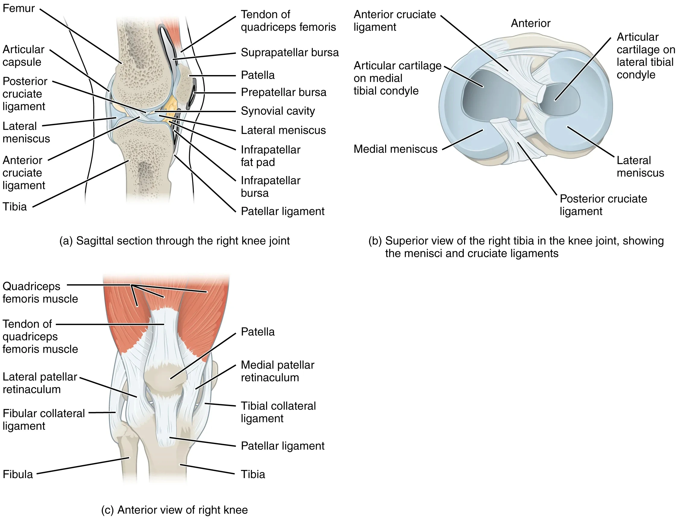
Labeled anatomical views of the knee showing bones, cartilage, menisci and major supporting ligaments.Open full-size diagram ↗
The knee jointOpenStax · source · CC BY 4.0

Explore Pilates movement connections ↗

Bone landmarks

The knee includes articulations between femur and tibia, and between femur and patella. Menisci help distribute load. The cruciate and collateral ligaments help guide and restrain movement; the fibula lies alongside the tibia.

Quadriceps
Straighten the knee; rectus femoris also flexes the hip.
Hamstrings
Bend the knee; most also extend the hip.
Popliteus
Helps initiate knee flexion from a locked position.

Connect to practice

Straight knees are not a measure of success in a forward fold. A bent-knee variation can change the task and make it easier to explore a comfortable hip hinge.

Adaptable yoga flows ↗
Back to body regions ↑

Region 10

Lower legs, ankles & feet

A mobile foundation from shin to toes.

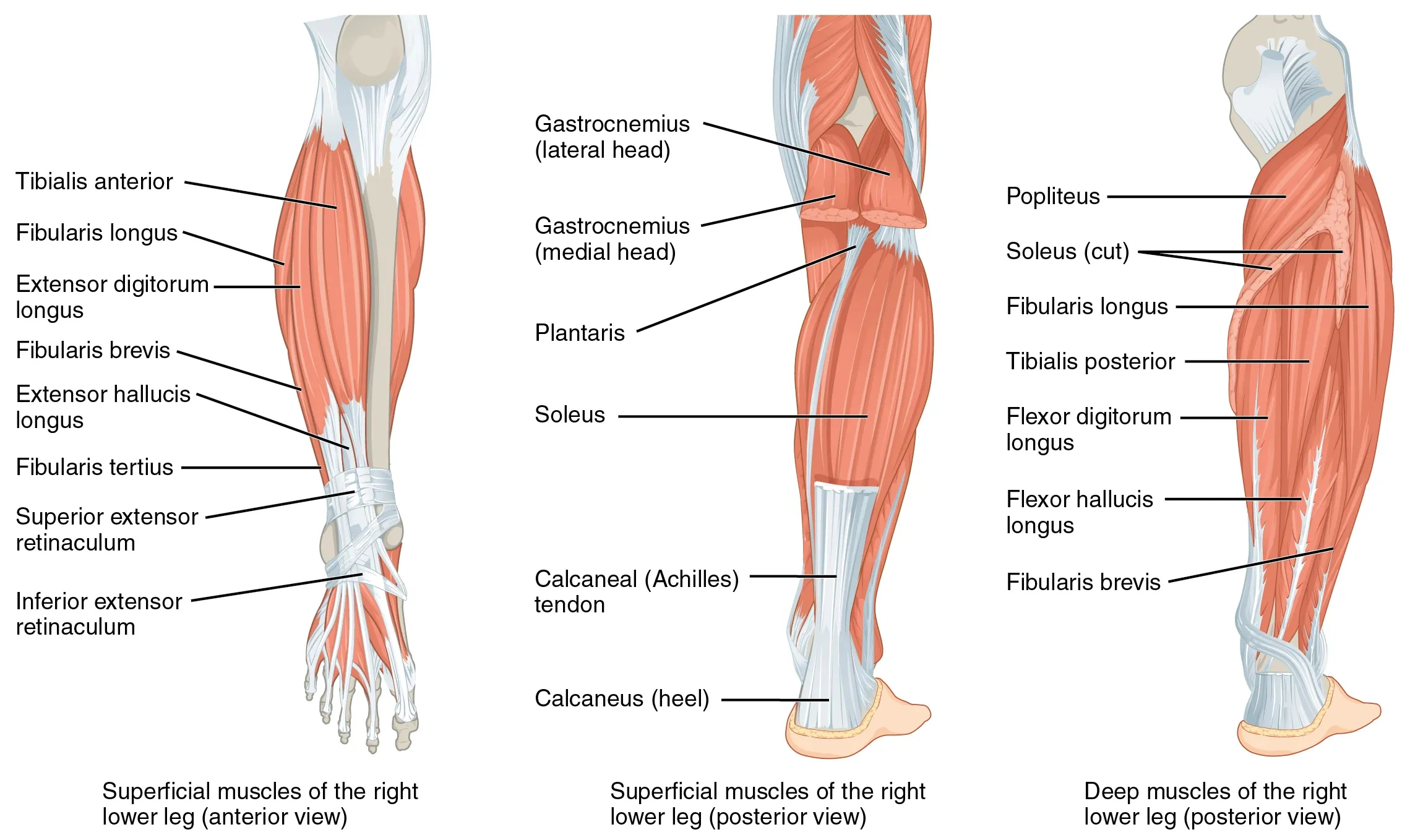
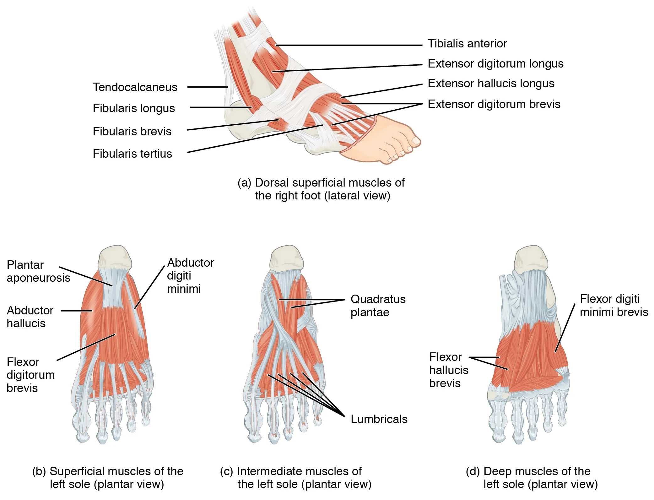
Labeled anterior and posterior views of lower-leg muscles, with superficial and deep posterior muscles including gastrocnemius, soleus and tibialis posterior.Open full-size diagram ↗
Muscles of the lower legOpenStax · source · CC BY 4.0

Explore Pilates movement connections ↗

Bone landmarks

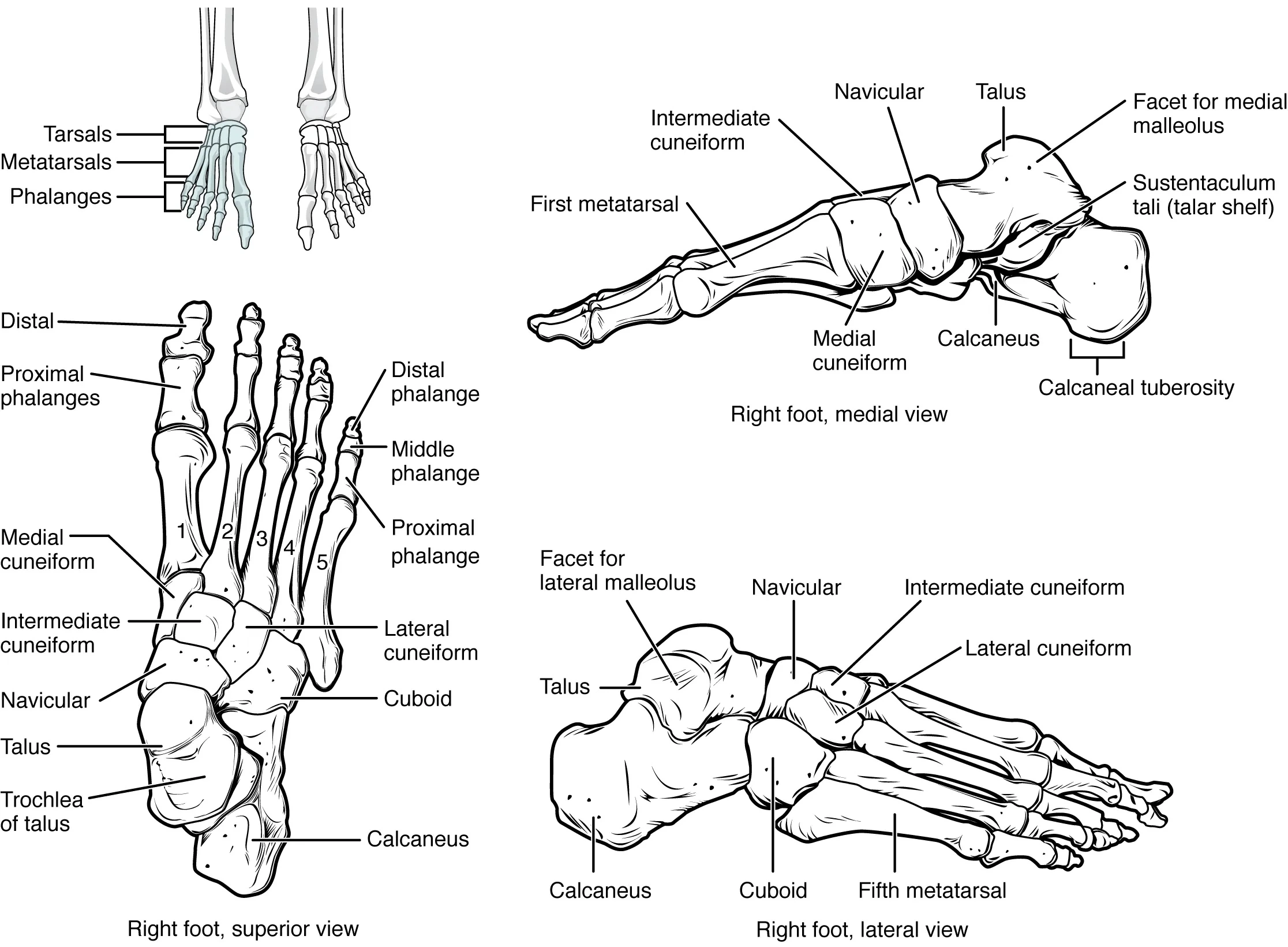
The tibia carries most lower-leg load; the fibula forms the outer ankle. Each foot has 7 tarsals, 5 metatarsals and 14 phalanges. The tarsals are the talus, calcaneus, navicular, cuboid and three cuneiforms.

Gastrocnemius & soleus
Raise the heel through the Achilles tendon.
Tibialis anterior
Lifts the front of the foot.
Tibialis posterior & fibularis muscles
Help control the foot and ankle.
Intrinsic foot muscles
Coordinate toes and contribute to arch support.
More diagrams · 2 plates

Connect to practice

Notice contact through the heel and the bases of the big and little toes in standing practice. A wall or chair can make balance exploration more accessible.

Standing practice ↗
Back to body regions ↑

04 · Muscle in focus

Trapezius: three fibre regions

This broad muscle runs from the back of the skull and midline of the neck and thorax to the collarbone and shoulder blade.

Anterior and posterior shoulder-girdle diagrams labeling trapezius, rhomboids, serratus anterior and other muscles that position the scapula.Open full-size diagram ↗
Muscles that position the shoulder girdleOpenStax · source · CC BY 4.0

The paired muscles form a broad, kite-like outline across the upper back. The regions work together; their effect depends on arm position and which structures are held steady.

Upper · descending fibres
Run from skull and neck towards the outer collarbone. Help elevate the shoulder girdle and contribute to the coordinated upward rotation used when raising the arm.
Middle · transverse fibres
Run across towards the acromion and scapular spine. Help draw the shoulder blade towards the spine and stabilise it.
Lower · ascending fibres
Run from the lower thoracic midline upwards to the scapular spine. Help depress and stabilise the scapula and contribute to upward rotation.

During an overhead reach, trapezius and serratus anterior coordinate shoulder-blade movement. The scapula rotates upward; this is different from simply shrugging the whole shoulder.

muscles of the pectoral girdle and upper limbs ↗

Focused study · lower leg

Calves: two muscles, different positions

The gastrocnemius and soleus help raise the heel through the Achilles tendon. Knee position changes which muscle is emphasised in a calf stretch.

Labeled anterior and posterior views of lower-leg muscles, with superficial and deep posterior muscles including gastrocnemius, soleus and tibialis posterior.Open full-size diagram ↗
Muscles of the lower legOpenStax · source · CC BY 4.0
Gastrocnemius · the superficial calf
Crosses the knee and ankle. A comfortable straight-knee wall stretch puts more emphasis on this muscle.
Soleus · the deeper calf
Crosses the ankle but not the knee. A small bent-knee wall stretch reduces gastrocnemius tension and places more emphasis on soleus.
Achilles tendon · the connection to the heel
Transmits force from the calf muscles to the calcaneus. A painful tendon or recent muscle strain is not a signal to stretch more strongly.

For mild tightness, begin with a supported wall variation rather than forcing the heels down in Downward Dog. Recurrent cramps, a recent pull and a swollen painful calf each need different guidance.

Open the finder and choose Calves & Achilles ↗

appendicular muscles of the pelvic girdle and lower limbs ↗Royal Orthopaedic Hospital: calf stretching ↗

Focused study · digestion

The digestive system

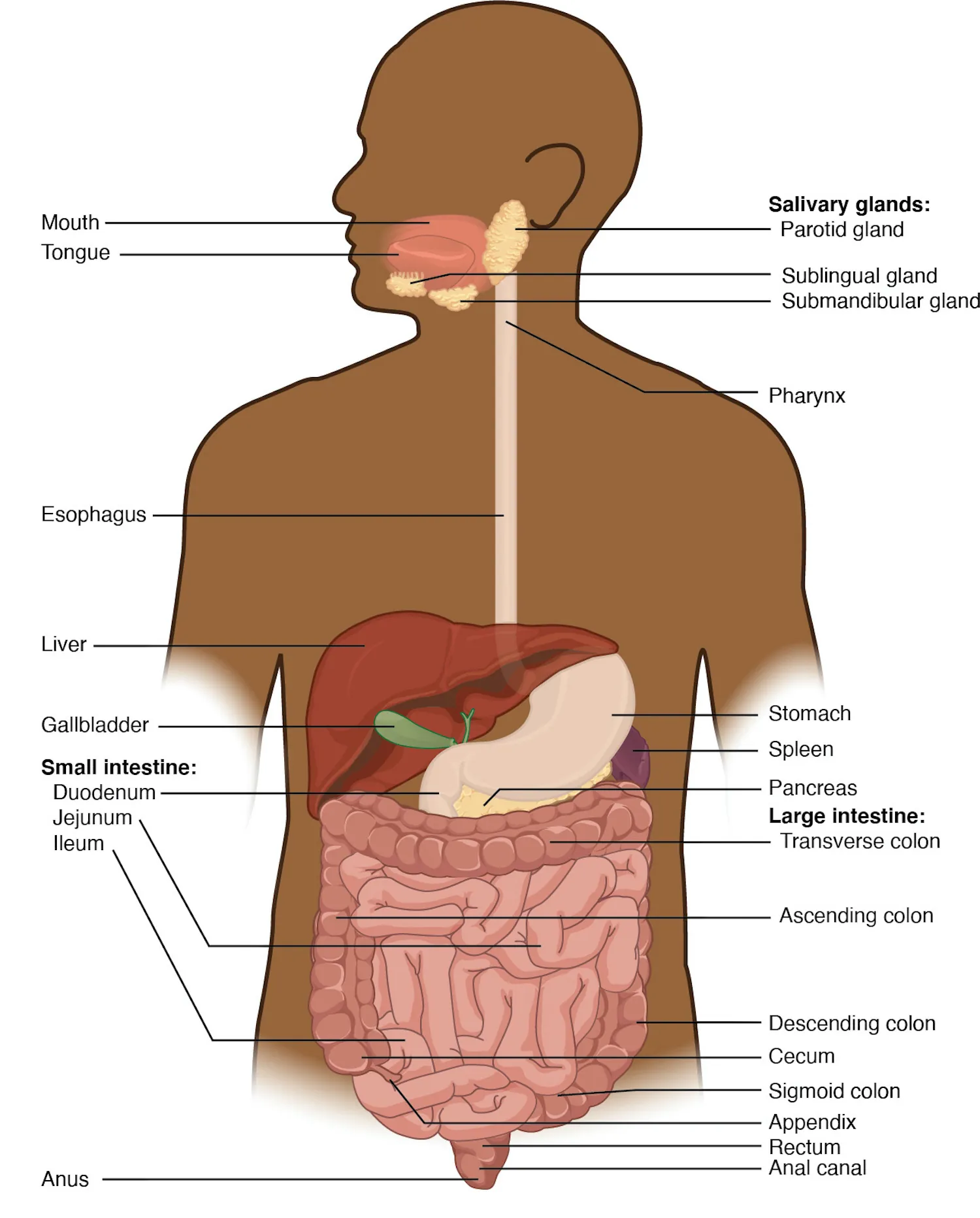
Food passes through the gut; accessory organs supply substances that help digestion. The abdominal muscles support this region but are not the bowel itself.

Labelled human digestive system showing mouth, tongue, salivary glands, pharynx, esophagus, liver, gallbladder, stomach, pancreas, small intestine, large intestine, appendix, rectum, anal canal, and anus. The nearby spleen is also labelled for orientation.Open full-size diagram ↗
Components of the Digestive SystemOpenStax · source · CC BY 4.0
Mouth, salivary glands & oesophagus
Chewing and saliva begin processing food. Swallowing moves it towards the stomach.
Stomach
Mixes food with acid and digestive secretions.
Small intestine
The duodenum, jejunum and ileum carry out much of digestion and nutrient absorption.
Liver, gallbladder & pancreas
The liver produces bile, the gallbladder stores it, and the pancreas supplies digestive enzymes and bicarbonate.
Large intestine, rectum & anus
Recover water, form and store stool, and allow its controlled passage.

Coordinated smooth-muscle activity moves and mixes gut contents. The spleen is shown for orientation; it belongs to the lymphatic and immune systems.

Movement and digestive comfort

Gentle activity and comfortable upright breathing can be supportive habits. Deep twists do not “detox” organs, and no stretch can diagnose or cure a digestive condition. New or persistent symptoms need appropriate assessment.

Explore digestive symptoms ↗

overview of the digestive system ↗NHS: bloating ↗NHS: constipation ↗

05 · Beyond muscles and bones

The body works together

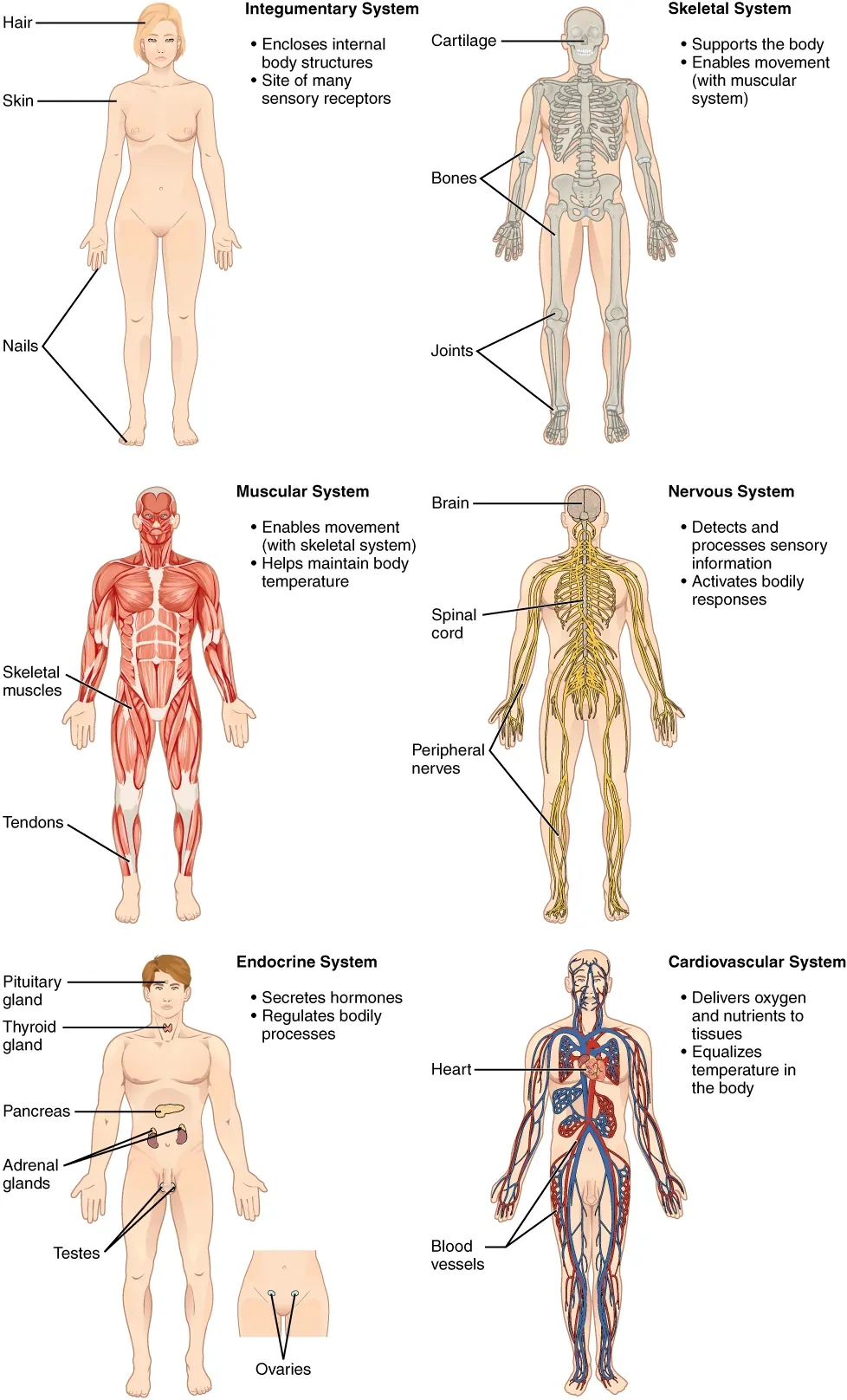
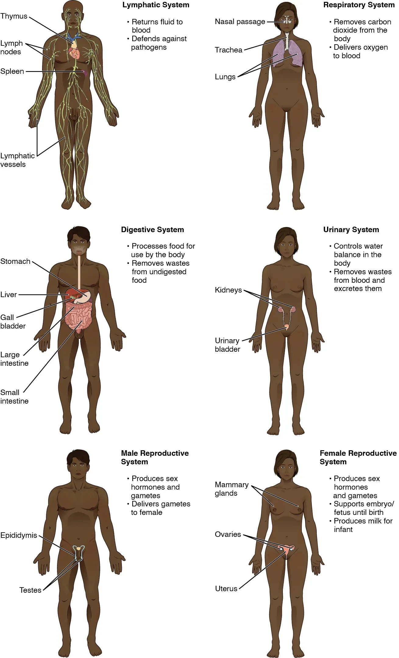
The anatomical systems are useful study categories. Their organs and functions are interdependent.

Integumentary

Skin, hair and nails

Protection and sensation.

Skeletal

Bones, cartilage and ligaments

Support, movement and protection.

Muscular

Skeletal muscles and tendons

Movement and heat production.

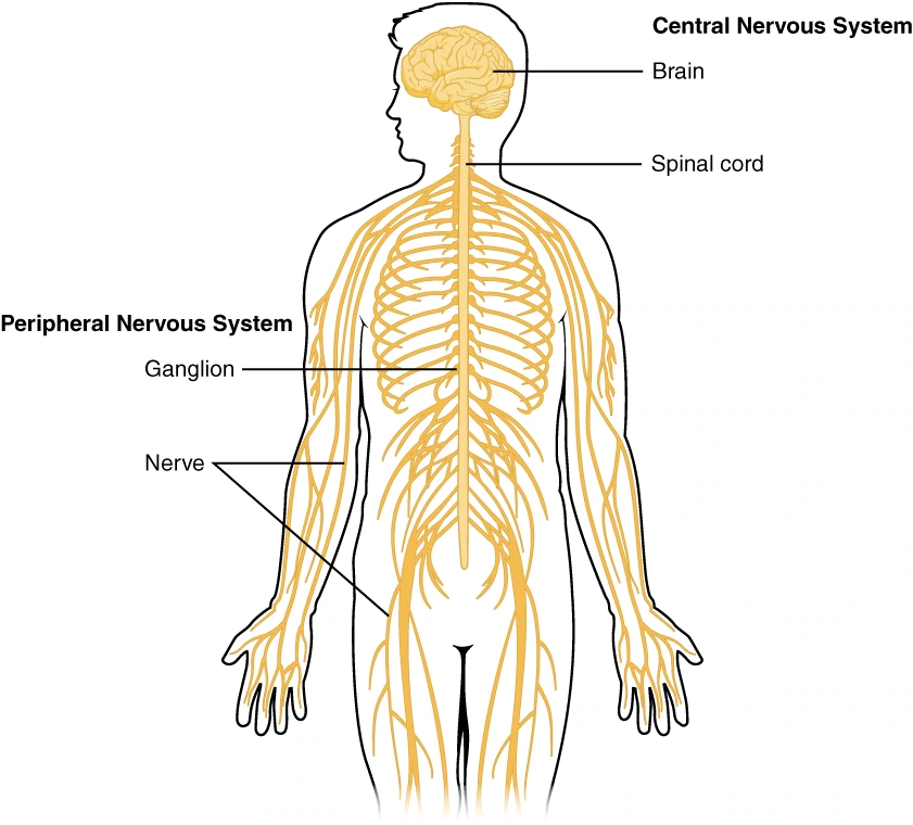
Nervous

Brain, spinal cord and nerves

Sensation, coordination and rapid communication.

Endocrine

Hormone-producing glands

Hormonal regulation.

Cardiovascular

Heart, blood and vessels

Transport of gases, nutrients and other substances.

Lymphatic & immune

Lymph vessels, nodes and related organs

Fluid return and immune defence.

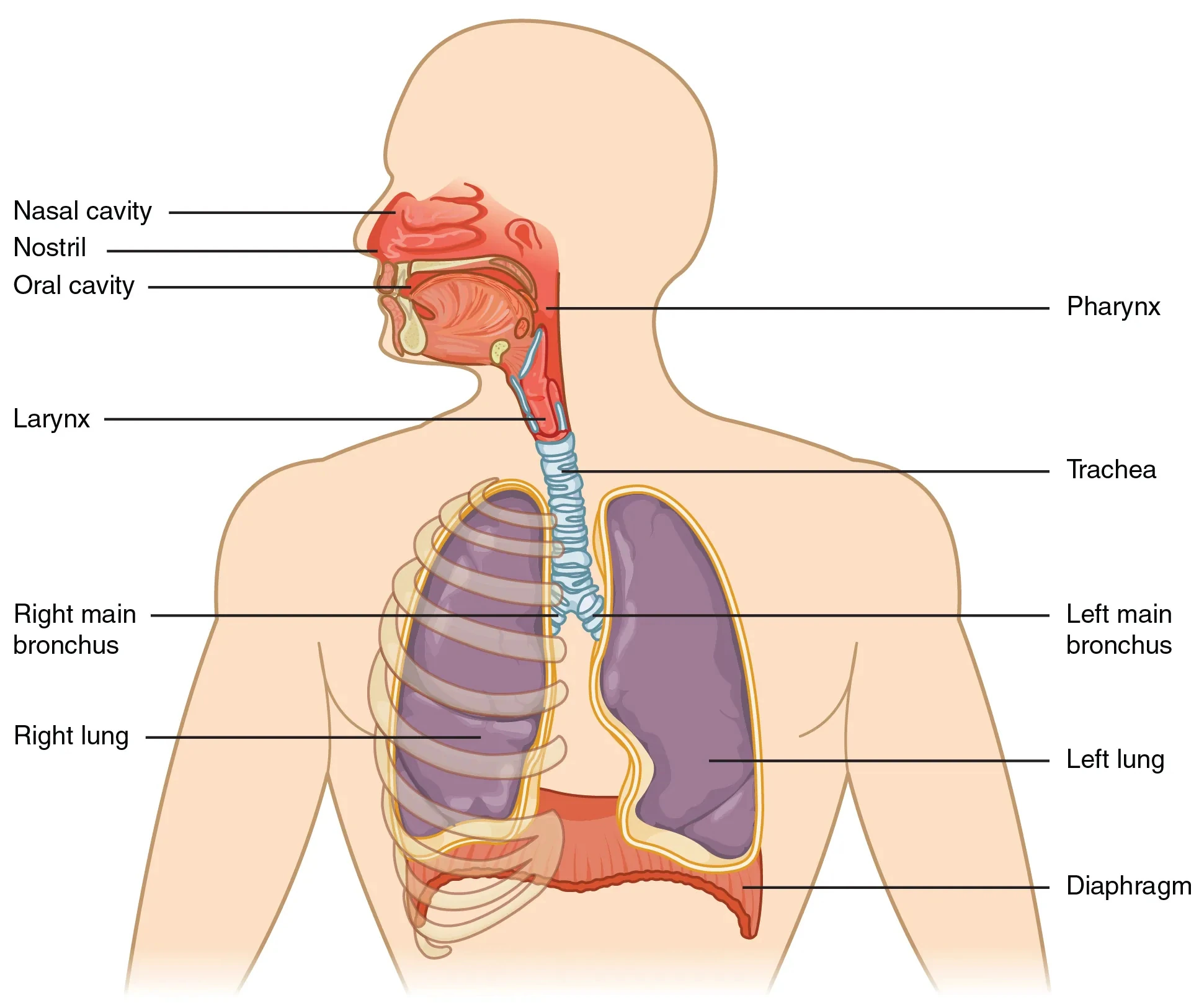
Respiratory

Airways and lungs

Exchange of oxygen and carbon dioxide.

Digestive

Digestive tract, liver and pancreas

Food processing and nutrient absorption.

Urinary

Kidneys, ureters and bladder

Waste excretion and fluid balance.

Reproductive

Reproductive organs

Reproduction and associated hormones.

Nervous and respiratory systems · closer views

structural organization of the human body ↗

06 · Read the language

What the terms mean

Use these definitions alongside the labels on the diagrams.

Flexion / extension
Bending / straightening a joint; at the trunk, bending forwards / backwards.
Abduction / adduction
Moving away from / towards the body’s midline. Fingers and toes use their own reference axes.
Internal / external rotation
Turning a limb around its long axis towards / away from the body’s midline.
Elevation / depression
Moving a structure up / down, as with the shoulder girdle.
Protraction / retraction
Moving the scapula around the ribs forwards / back towards the spine.
Dorsiflexion / plantar flexion
Bringing the foot towards the shin / pointing it away.
Pronation / supination
Turning the forearm so the palm faces down / up when the elbow is bent.
Anterior / posterior
Front / back. Medial means nearer the midline; lateral means farther from it.

types of body movements ↗

Sources & illustration credits

Illustrations: OpenStax, Anatomy and Physiology, from the original textbook and Wikimedia Commons. Each plate links to its source and Creative Commons licence. Image format and size are adapted for the web; anatomical content and labels are unchanged.

Access the textbook for free at OpenStax ↗

House of Ananda’s practice connections are educational examples. The atlas covers major structures across the body; it is not an exhaustive catalogue of every muscle or a diagnosis of an individual body.

Back to the atlas ↑

From Daniel’s anatomy notebook

Study the systems. Learn the bone counts.

Two connected lessons explain cells, tissues, organs, the skeleton, limb bones and girdles, with illustrated reference tables.

Body organisation & systems ↗Bones, limbs & girdles ↗

Ten systems and regions

From breath
to ground.

A yoga-informed overview without pretending every body should create the same pose.

01

Breath & diaphragm

The diaphragm is the principal muscle of quiet breathing. Rib movement, abdominal pressure and pelvic-floor response all contribute.

In practice

Let the breath change shape naturally. Bigger is not always better, and breath-holding is a separate advanced skill.

02

Spine

The cervical, thoracic and lumbar regions have different structures and typical ranges. The spine shares movement across many joints.

In practice

Create length before loading rotation. Backbends need thoracic movement, hip extension and trunk support - not only lumbar compression.

03

Shoulder complex

The upper arm, shoulder blade, collarbone and ribcage work as a coordinated system rather than one isolated joint.

In practice

Allow shoulder blades to move. In weight-bearing poses, distribute load through hands, arms and upper back.

04

Wrists & hands

The wrist contains multiple small bones and joints. Finger pressure and forearm strength help share load when the hands bear weight.

In practice

Progress loading gradually. Reduce angle with wedges, use fists or forearms, and stop with sharp pain or numbness.

05

Pelvis & core

The pelvis transfers force between trunk and legs. The abdominal wall, back muscles, diaphragm and pelvic floor coordinate pressure and movement.

In practice

Core work is not constant bracing. Use enough support for the task while preserving breath and adaptability.

06

Hips

The ball-and-socket hip allows movement in several planes, but bone shape differs substantially between people.

In practice

There is no universal perfect depth. Respect compression and avoid forcing the knee to manufacture hip rotation.

07

Knees

The knee primarily bends and straightens, with smaller rotational movement. Ligaments, menisci and surrounding muscles guide stability.

In practice

Track the knee in a comfortable direction and adjust stance width. Pain is information, not a flexibility challenge.

08

Feet & ankles

The foot adapts to the ground through many joints. Calf strength and ankle mobility influence squats, lunges and balance.

In practice

Use the whole foot rather than gripping the toes. Heel support is a valid tool when ankle range limits a squat.

09

Fascia & mobility

Connective tissues surround and link structures, but mobility also depends on nervous-system tolerance, joint shape, strength and practice history.

In practice

Avoid claims that one pose 'releases' all fascia. Combine varied movement, progressive loading and recovery.

10

Nervous system

Balance, coordination, pain and effort are interpreted by the nervous system. Sensation is influenced by context, stress, sleep and previous experience.

In practice

Slow repetition, choice and predictable cues can support learning. Pain does not always equal damage, but persistent pain deserves assessment.
Myth and reality

Clearer language.
Safer practice.

Perfect alignment prevents injury

Alignment is useful, but bodies and tasks vary. Capacity, load, history, recovery and context also matter.

Tight muscles simply need stretching

A feeling of tightness can reflect many factors. Strength, graded exposure, rest or clinical assessment may be more useful.

More flexibility means more advanced yoga

Yoga includes ethics, breath, concentration and discernment. Extreme range is neither necessary nor universally safe.

Twists detox the organs

Twists create movement and pressure changes, but the liver, kidneys, lungs and digestive system perform the body's metabolic processing.

When to pause

Sensation needs
interpretation.

Stop and seek appropriate advice for new severe pain, trauma, marked swelling, loss of strength, numbness, fainting, chest pain, visual disturbance or symptoms that persist or worsen. A yoga teacher does not diagnose injury unless separately qualified to do so.

Practice should be adapted for pregnancy, recent surgery, joint replacement, osteoporosis, cardiovascular conditions, glaucoma and other relevant health factors.

NCCIH: yoga effectiveness and safety ↗

Connected study · emotions and the body

Explore emotion, muscle tension and gentle practice.

Read the illustrated thesis, explore the regional muscle atlas, and try twelve gentle practices with clear instructions and adaptations.

Enter Emotions, Muscles & the Body ↗