 House of AnandaMovement · Stillness · Wisdom · Journey
Lesson 10 of 14 · Yoga & anatomy
Joints & movement
Where bones meet—and movement becomes possible.
Structural and functional joint categories, the synovial-joint sketch, skull sutures, spinal joints and the bone–cartilage distinction.
01 · Study
Not every joint has a fluid cavity
A joint, or articulation, is a meeting between skeletal elements. Joints differ in structure and movement. Fibrous, cartilaginous and synovial are structural categories; the fluid-filled sketch represents the synovial type.
02 · Study
Read the four labels
Ligaments connect bone to bone and contribute stability. Tendons link muscles to bones; muscular activity also helps control a joint. The simple sketch leaves out these additional structures.
| Structure | Where / what it does |
|---|---|
| Articular cartilage | Smooth covering on the articulating bone ends; reduces friction. |
| Joint capsule | Encloses the joint; its outer fibrous layer provides support. |
| Synovial membrane | Inner lining that produces synovial fluid; does not cover the articular cartilage. |
| Synovial fluid | Lubricates the joint cavity and helps nourish cartilage. |
Further reading: OpenStax · Synovial joints ↗
03 · Study
Structure and mobility are different classifications
The notebook’s “perfect movable” means freely movable, not unlimited movement. Fibrous, cartilaginous and synovial classify structure; synarthrosis, amphiarthrosis and diarthrosis classify function.
Sutures join skull bones without a synovial cavity. The spine also includes synovial facet joints, so “spinal joints have no cavity” is too broad. Rib connections are not all one type.
| Functional name | Movement | Example |
|---|---|---|
| Synarthrosis | Essentially immovable | Most adult skull sutures. |
| Amphiarthrosis | Slightly movable | Joints between adjacent vertebral bodies, joined by discs. |
| Diarthrosis | Freely movable | Synovial joints, such as hip or shoulder. |
Further reading: OpenStax · Classification of joints ↗
04 · Study
A healing comparison, not an absolute
The note says bones heal themselves but teeth cannot. Bone has repair capacity, although a fracture may need treatment and does not always unite normally. Adult tooth enamel does not regrow like bone; early mineral loss can sometimes be remineralised. Teeth also contain living tissues, so the blanket claim needs qualification.
Cartilage is not a universal “filler” in every joint. The synovial-joint diagram above and the disc lesson describe different arrangements. None of these comparisons can diagnose an injury from symptoms alone.
Further reading: NIDCR · The tooth decay process ↗
05 · Study
Bring anatomy into practice
Joint names describe structures; they do not prescribe one ideal depth for every posture. The notes are a foundation for understanding instructions, not for diagnosing pain. Use the anatomy atlas to place the diagram in the whole body.
Further reading: NCCIH · Yoga effectiveness and safety ↗
06 · Study
Six shapes of synovial joint
“Freely movable” does not mean unrestricted. A hinge primarily moves around one axis, not in only one direction.
| Type | Typical movement | Example |
|---|---|---|
| Plane / gliding | Small sliding movements | Some intercarpal joints. |
| Hinge | Mainly bending and straightening | Elbow humeroulnar joint. |
| Pivot | Rotation around an axis | Proximal radioulnar joint. |
| Condyloid / ellipsoid | Movement in two principal planes | Wrist radiocarpal joint. |
| Saddle | Two-plane movement with opposing saddle surfaces | Thumb carpometacarpal joint. |
| Ball-and-socket | Movement around several axes | Hip and shoulder. |
Further reading: OpenStax · Synovial joints ↗
07 · Study
Bursae and the moving ribcage
A bursa is a small fluid-containing sac that reduces friction between neighbouring tissues. It is not another name for the whole synovial joint or its cavity.
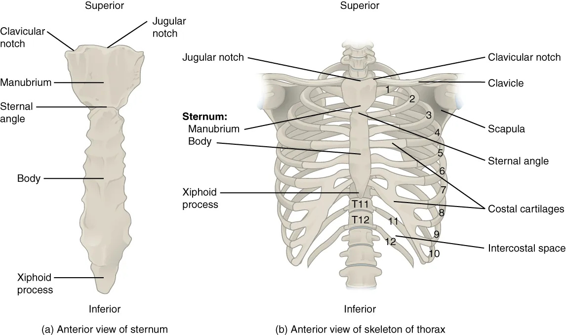
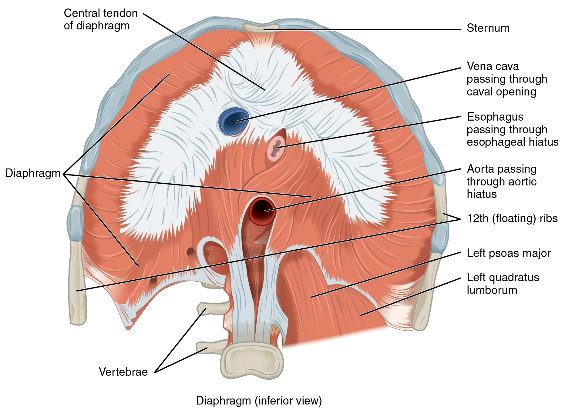
The ribcage moves with breathing through coordinated joint movement, cartilage deformation and respiratory-muscle activity. It is too simple to class all rib connections as slightly movable cartilaginous joints.

Further reading: OpenStax · Synovial joints ↗
From notebook to page
IMG_1590, left page. The drawing shows the basic components of a synovial joint rather than identifying a particular knee, hip or other joint. Further study: IMG_1858: functional joint categories and skull/spine examples. Further study: IMG_1859: ribs, bursae and six synovial joint types.
Spelling has been standardised and explanations added. Traditional teaching cues, anatomical facts and uncertain handwriting are distinguished in the text. These pages support learning alongside appropriate teaching.