Movement / Pilates / Pilates & anatomy Foundations Exercise library Guided sessions Programmes History & principles Classical 34 Pilates & anatomy Pilates & yoga Equipment studio Teachers & sources The muscles work as coordinated groups. Highlighting a region explains a movement focus; it does not isolate a single muscle or establish treatment for a painful area.
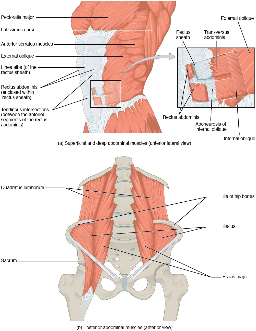
OpenStax · source · CC BY 4.0 Abdomen, pelvis & pelvic floor Support, pressure and movement through the centre.
Rectus abdominis Flexes the trunk.
Internal & external obliques Contribute to trunk rotation and side bending.
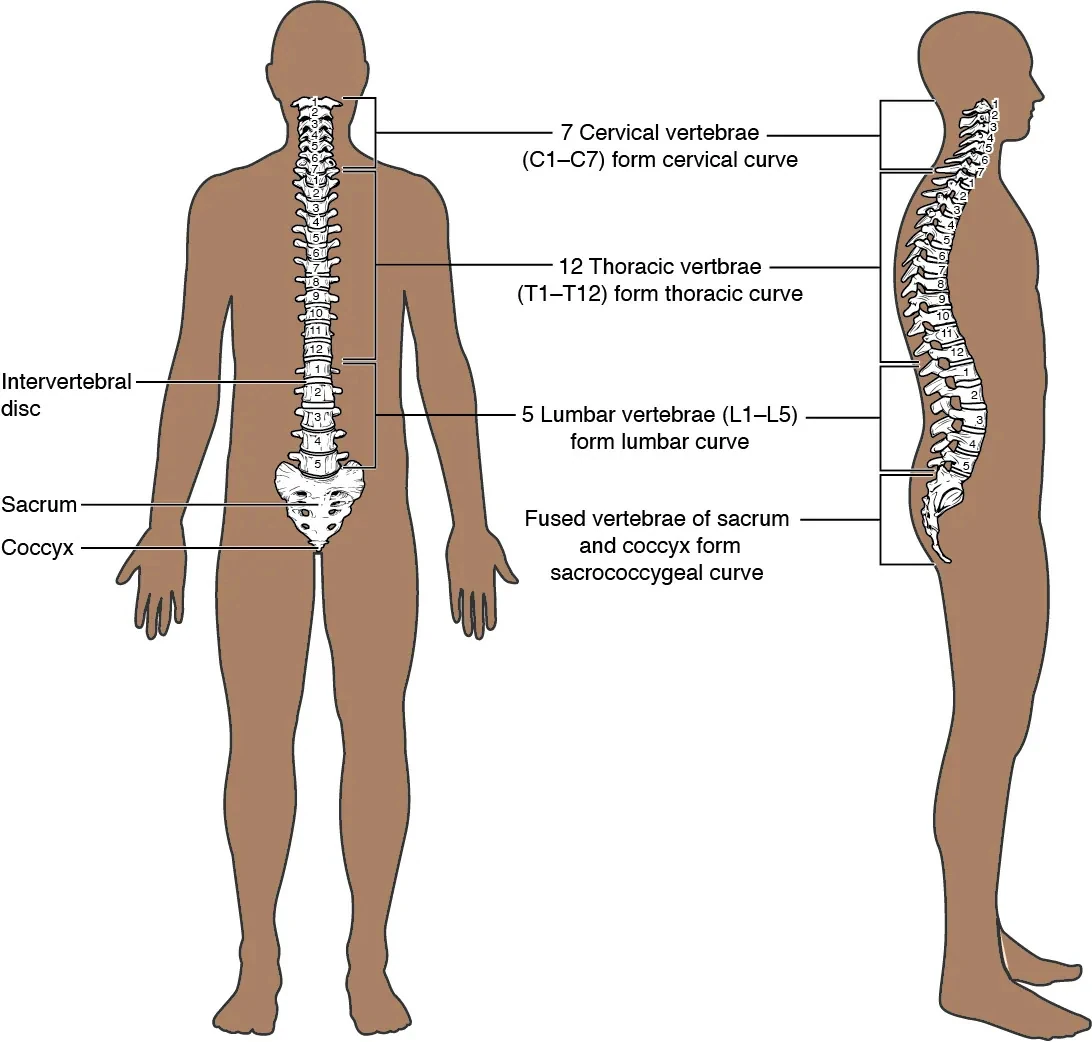
Transversus abdominis Contributes to abdominal compression and trunk support. OpenStax · source · CC BY 4.0 Spine & back A column of joints, supported by layers of muscle.
Erector spinae Extend the spine and help control side bending.
Multifidus & other deep spinal muscles Help control movement between spinal segments.
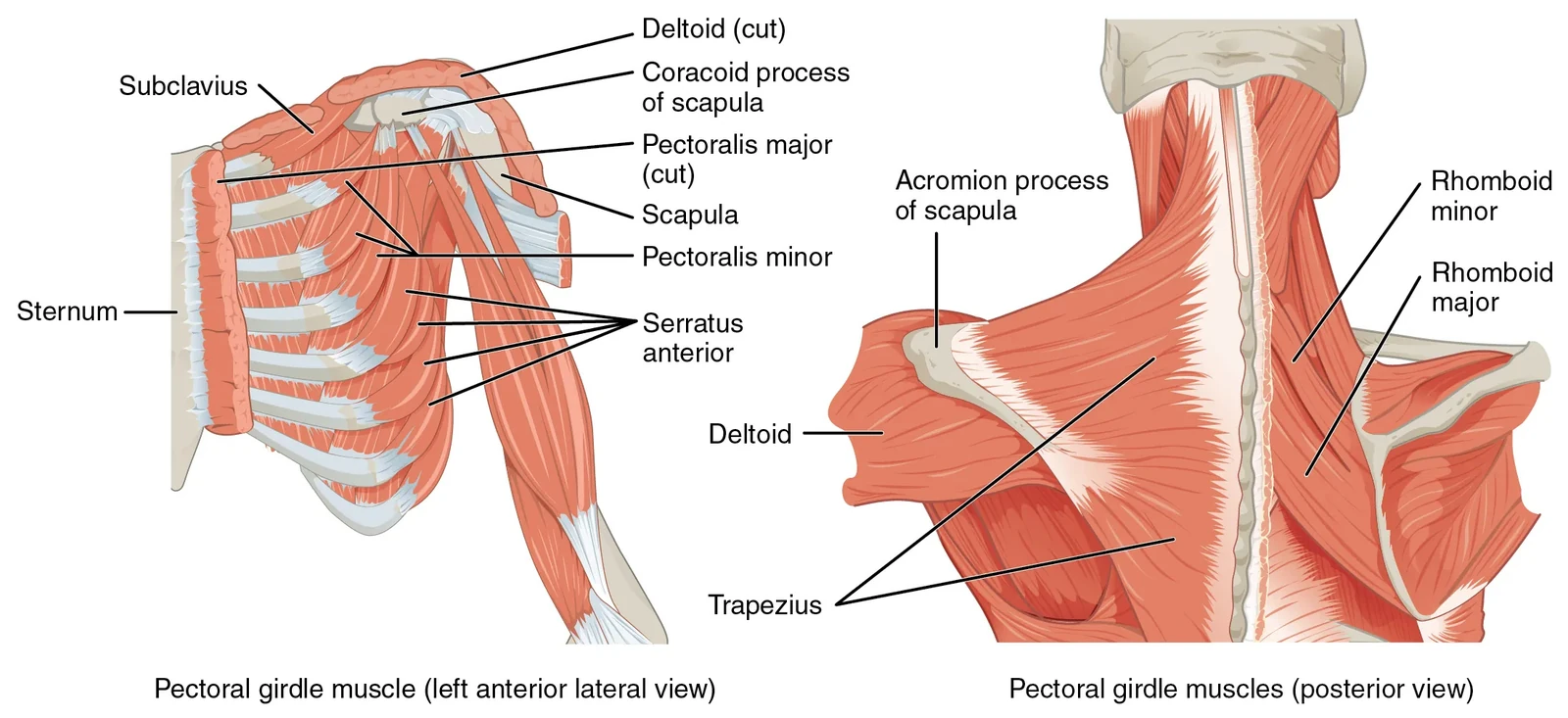
Quadratus lumborum Assists side bending and supports the twelfth rib. OpenStax · source · CC BY 4.0 Shoulders & upper back The arm, shoulder blade and collarbone move together.
Trapezius Positions and stabilises the shoulder blade; its fibre groups contribute differently.
Serratus anterior Draws the scapula around the ribs and helps it rotate upward.
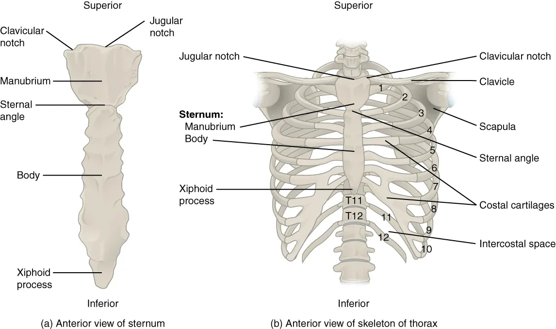
Rhomboids Draw the scapula towards the spine. OpenStax · source · CC BY 4.0 Chest, ribs & breathing A moving cage around the heart and lungs.
Diaphragm Contracts and descends during inhalation, increasing chest-cavity volume.
Intercostals Muscles between the ribs assist breathing mechanics.
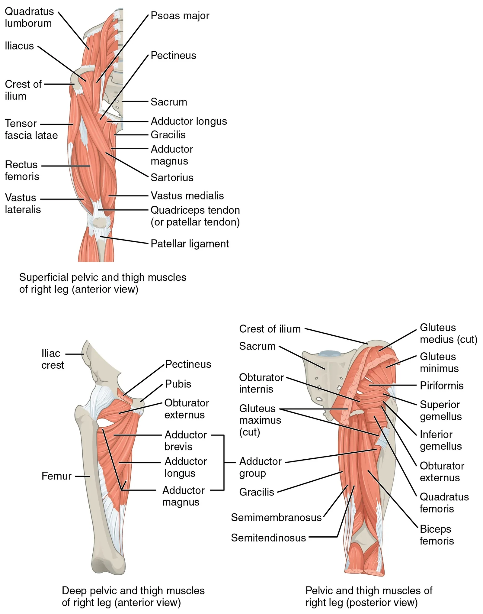
Pectoralis major Moves the upper arm across and towards the body. OpenStax · source · CC BY 4.0 Hips, buttocks & thighs Large muscles move the femur and steady the pelvis.
Iliopsoas Psoas major and iliacus flex the hip.
Gluteal muscles Gluteus maximus extends the hip; medius and minimus assist pelvic control and hip abduction.
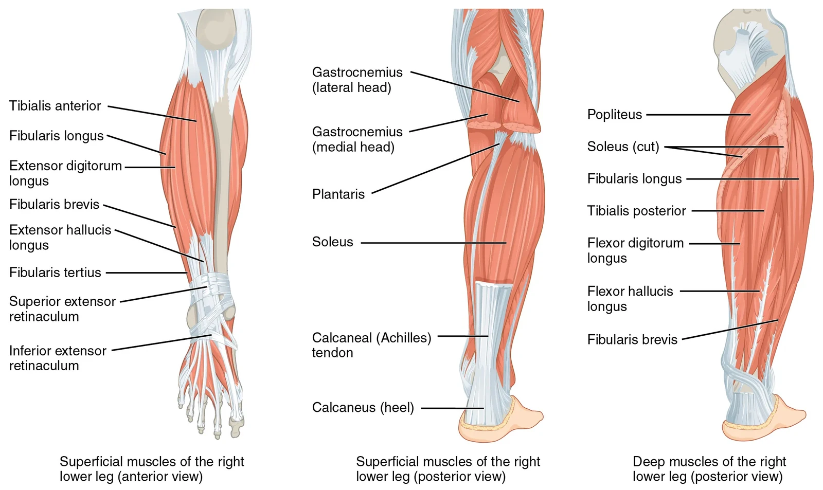
Adductors Draw the thigh towards the midline. OpenStax · source · CC BY 4.0 Lower legs, ankles & feet A mobile foundation from shin to toes.
Gastrocnemius & soleus Raise the heel through the Achilles tendon.
Tibialis anterior Lifts the front of the foot.
Tibialis posterior & fibularis muscles Help control the foot and ankle. Calves, breathing and the wider body Use supported heel raises to study calf loading, and rib breathing to notice respiratory movement. The digestive organs and abdominal muscles have different roles; trunk exercises are not treatment for digestive disease.
Scope & review
Learn with clear limits. Prepared for House of Ananda with AI assistance. These original exercise descriptions, diagrams and programme drafts have not yet been reviewed by a named qualified Pilates teacher. They are general education for adults comfortable with the positions shown.
Stop for pain, dizziness or feeling unwell. Ask a qualified professional about injury, unexplained symptoms, pregnancy, recent surgery or other relevant health conditions before following a session. Choose comfortable alternatives and pause freely.
Teaching, sources and review details → Editorial release: 12 September 2026 · Qualified teacher review: pending.