 House of AnandaMovement · Stillness · Wisdom · Journey
Lesson 13 of 14 · Yoga & anatomy
Muscles, contraction & movement
Understand the work beneath the shape.
Three muscle tissues, contraction types, muscle roles and the neck, back, hip, hamstring and calf names in the notebook.
01 · Study
Three muscle tissues
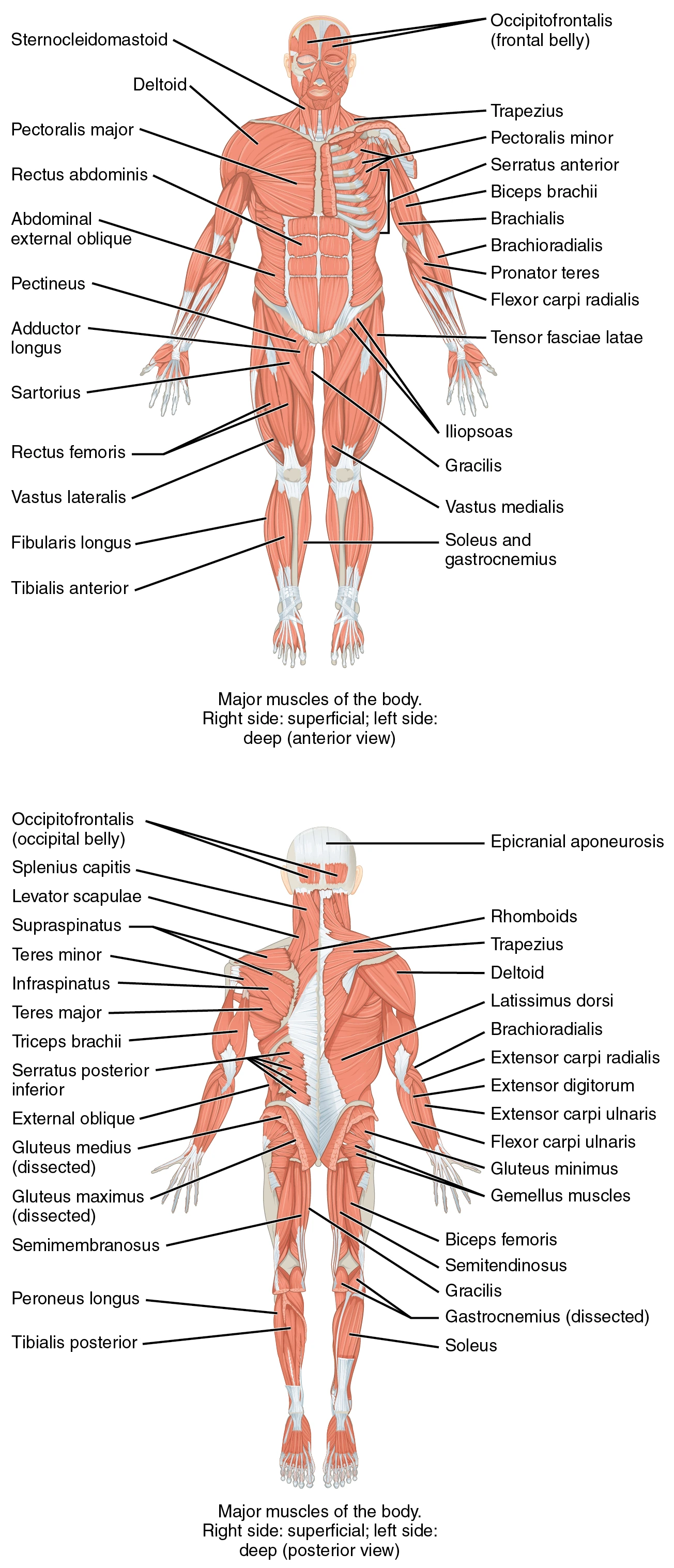
The notebook’s “600 muscles” is an approximate conventional count of skeletal muscles, not a total of all muscle cells. Muscle activity supports movement, posture, joint stability and heat production. Breathing is not performed exclusively by smooth muscle: the diaphragm is skeletal muscle.
| Tissue | Typical location | Control |
|---|---|---|
| Skeletal | Most muscles moving the skeleton; also the diaphragm | Voluntary actions, with reflex and automatic contributions. |
| Cardiac | Heart wall | Involuntary rhythmic activity. |
| Smooth | Walls of hollow organs and blood vessels | Largely involuntary. |

Further reading: OpenStax · Muscle tissues ↗
02 · Study
Properties are not training goals
Extensibility does not mean a muscle should be pushed as far as possible. A sensation of tightness alone cannot establish which tissue is limiting movement.
| Term | Meaning |
|---|---|
| Excitability | Responding to a stimulus; “irritability” is an older physiological synonym, not an emotion. |
| Contractility | Generating force. |
| Extensibility | Being lengthened within a physiological range. |
| Elasticity | Recoil towards an earlier length. |
| Adaptability | A broader response to repeated demands, rather than a separate basic tissue property in every textbook. |
Further reading: OpenStax · Muscle tissues ↗ · NCCIH · Yoga effectiveness and safety ↗
03 · Study
A contraction does not always shorten a muscle
Isometric does not mean “strength” while isotonic means “flexibility”: both can involve substantial effort. A pose combines different actions across muscles. Keep breathing rather than treating a hold as a breath-holding exercise.
| Action | What changes | Study example |
|---|---|---|
| Concentric | Active muscle shortens | Elbow flexors lifting a light object. |
| Eccentric | Active muscle lengthens while controlling a load | The same flexors lowering it slowly. |
| Isometric | Force with little change in overall muscle length or joint angle | Holding the object still. |
| Isotonic | Length changes against a broadly steady load; an idealised term | Often used to group concentric and eccentric actions. |
| Isokinetic | Movement at a controlled constant angular speed | Usually studied using specialised equipment. |
Further reading: OpenStax · Control of muscle tension ↗
04 · Study
Agonist, antagonist and synergist
These are changing roles, not permanent labels on a muscle. The antagonist is not simply switched off: muscles may co-contract to control a joint. The notebook’s “atrophy” means loss of muscle size; it cannot be identified from feeling stiff or tired.
| Role | In a particular action |
|---|---|
| Agonist | A main contributor to the intended movement. |
| Antagonist | Can oppose or help control that movement. |
| Synergist | Assists or stabilises so the action can occur. |
05 · Study
Neck, shoulder blade and back
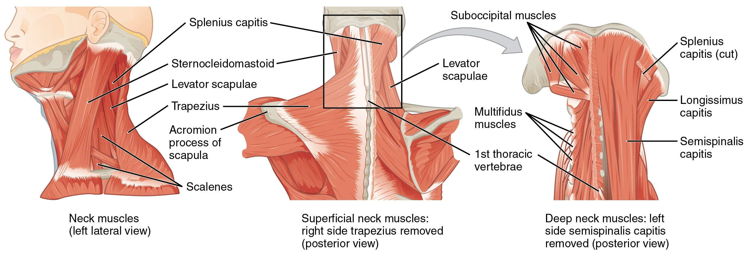
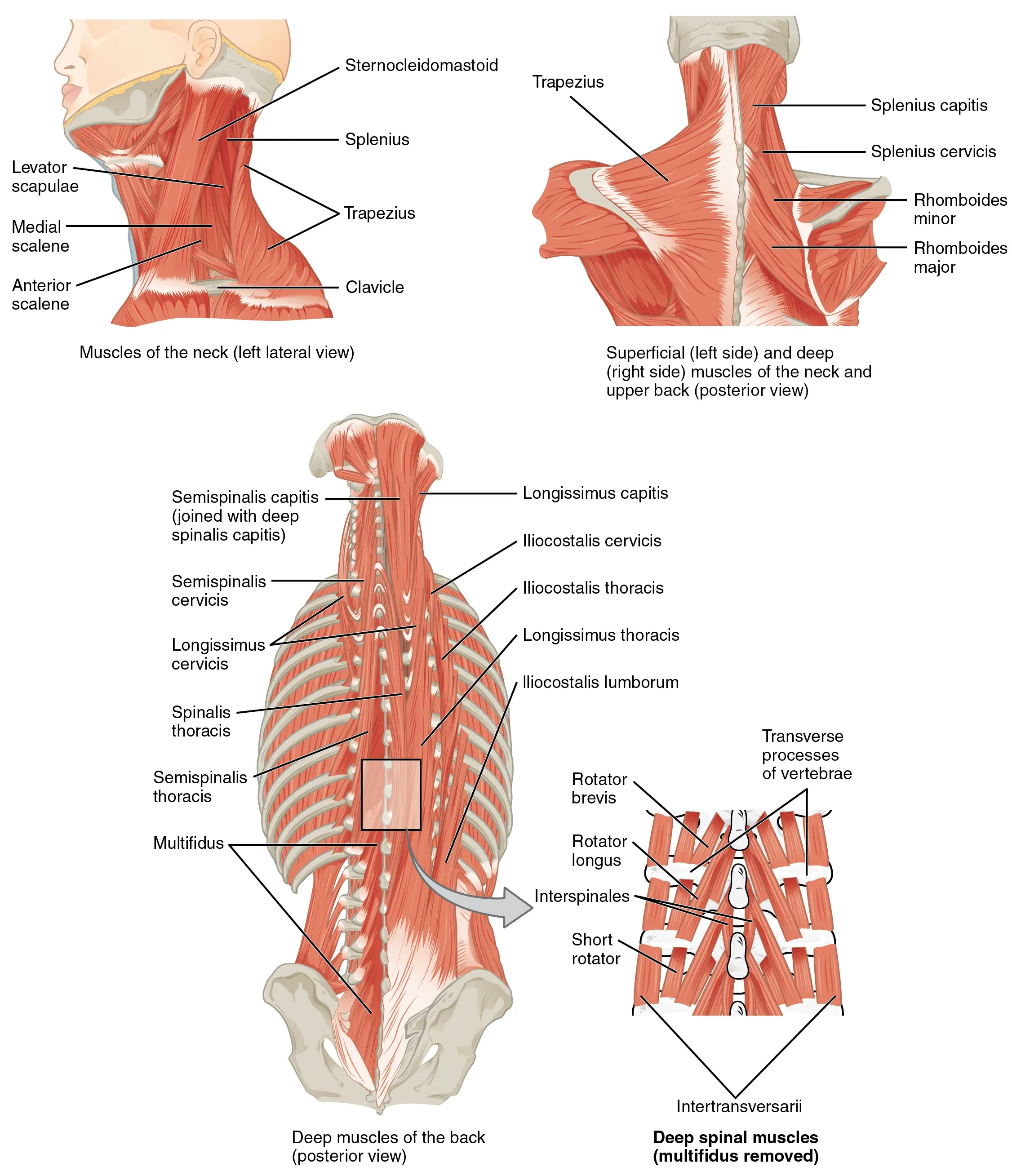
Torticollis is a clinical term for an abnormal neck posture with several possible causes, not a synonym for ordinary stiffness. Do not force the head into position from the notebook’s “wry neck” annotation.
| Name | Location and action | Notebook clarification |
|---|---|---|
| Sternocleidomastoid | Front/side of neck; contributes to turning and bending the head | Not a diagnosis of every stiff neck. |
| Levator scapulae | Neck to upper inner shoulder blade; helps elevate the scapula | Neck side-bending depends on what is fixed. |
| Trapezius | Broad superficial upper-back muscle; portions move and stabilise the scapula differently | The two sides form the familiar broad outline. |
| Rhomboid major and minor | Deep to trapezius; retract and stabilise the shoulder blade | C7–T5 is a combined regional shorthand, not one attachment for both muscles. |
| Latissimus dorsi | Broad back muscle acting on the upper arm | Helps extend, adduct and internally rotate the humerus. |


Further reading: OpenStax · Head, neck and back ↗ · OpenStax · Shoulder girdle and upper limb ↗
06 · Study
Hips, hamstrings and calves
Gluteus maximus is not a fourth hamstring. “Plantar flexion” means pointing the foot away from the shin, not folding the trunk. Muscles that cooperate in a movement need not belong to the same anatomical group.
| Group | Main study points |
|---|---|
| Iliopsoas | Iliacus and psoas major work together in hip flexion. |
| Gluteus maximus | A major hip extensor. |
| Gluteus medius and minimus | Contribute to hip abduction and pelvic control. |
| Hamstrings | Biceps femoris, semitendinosus and semimembranosus; bend the knee, with most portions also extending the hip. |
| Gastrocnemius and soleus | The major calf muscles; both plantar-flex the ankle. Gastrocnemius also crosses the knee. |
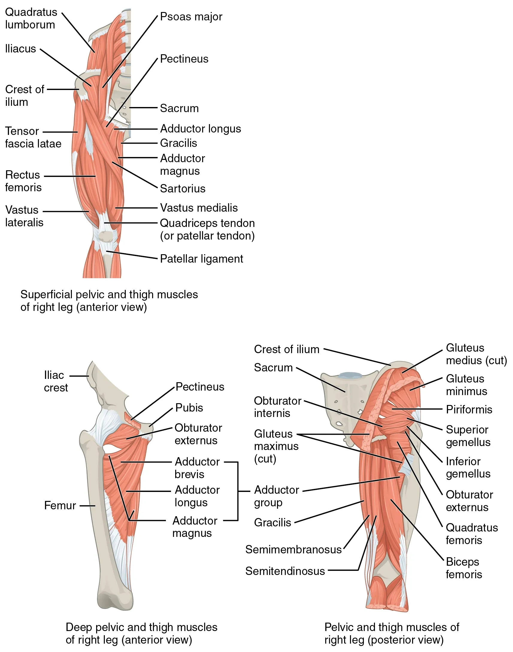
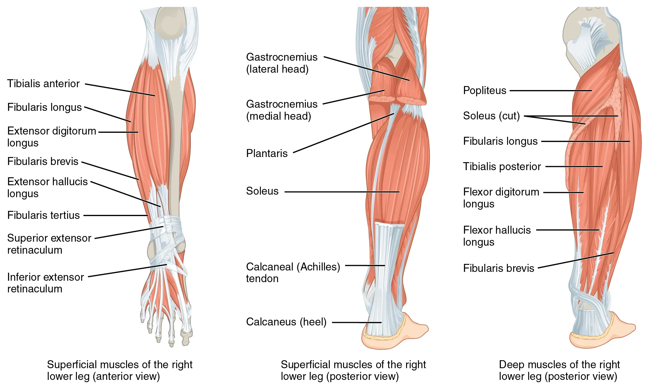
Further reading: OpenStax · Pelvic girdle and lower limb ↗
07 · Study
Use the map without self-diagnosing
The class ends with “strengthening and stretch”. These are complementary possibilities, not an instruction to stretch every painful area. Range, load, recovery and control all matter; an illustrated muscle cannot tell you the cause of a symptom.
Further reading: NCCIH · Yoga effectiveness and safety ↗
From notebook to page
IMG_1859–1860, IMG_1866 and IMG_1868–1869. Spellings are standardised; incomplete attachment labels and clinical shorthand are qualified rather than turned into diagnoses.
Spelling has been standardised and explanations added. Traditional teaching cues, anatomical facts and uncertain handwriting are distinguished in the text. These pages support learning alongside appropriate teaching.